 Journal of Medical Research and Surgery
PROVIDES A UNIQUE PLATFORM TO PUBLISH ORIGINAL RESEARCH AND REMODEL THE KNOWLEDGE IN THE AREA OF MEDICAL AND SURGERY
 Journal of Medical Research and Surgery
PROVIDES A UNIQUE PLATFORM TO PUBLISH ORIGINAL RESEARCH AND REMODEL THE KNOWLEDGE IN THE AREA OF MEDICAL AND SURGERY
 Indexed Articles
Select your language of interest to view the total content in your interested language
Helder Santos1*, Hugo Miranda1, Inês Almeida1, Mariana Santos1, Joana Chin1, Lurdes Almeida1
1Cardiology Department, Centro Hospitalar Barreiro Montijo E.P.E, Barreiro, Portugal.
Correspondence to: Helder Santos, Cardiology Department, Centro Hospitalar Barreiro Montijo E.P.E, Barreiro, Portugal; E-mail: helder33689@gmail.com
Received date: July 21, 2020; Accepted date: July 29, 2020; Published date: August 5, 2020
Citation: Santos H, Miranda H, Almeida I , et al. (2020) Two precious points in Cardiology. J Med Res Surg. 1(4): pp. 1-2.
Copyright: ©2020 Santos H,et al. This is an open-access article distributed under the terms of the Creative Commons Attribution License, which permits unrestricted use, distribution and reproduction in any medium, provided the original author and source are credited.
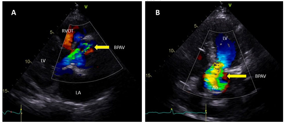
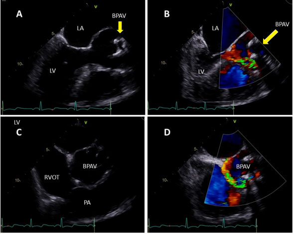
LA: Left Atrium; LV: left Ventricle; RVOT: Right Ventricular Outflow Tract, BPAV: Bio-Prosthesis Aortic Valve, Ao: Aorta; PA: Pulmonary Artery
A 74 year-old women with medical history of aortic replacement for severe aortic stenosis with a bio-prosthesis six years before and chronical kidney disease. Presented in the cardiology outpatient clinic complaining of dyspnea to minimal exertion, in class II, until them always in class I NYHA. She underwent a Transthoracic Echocardiogram (TTE), which showed a moderate periprosthetic leak, suggesting an anterior prosthetic valve dehiscence (Figure 1).
 Figure 1: Transthoracic echocardiogram; (A) Parasternal long-axis view showing a periprosthetic leak; (B) Apical 5 chambers view demonstrating a moderate
periprosthetic leak.
 Progressively increasing shortness of breath, paroxysmal nocturnal dyspnea, fatigue, being performed 4 months later a Transesophageal Echocardiogram (TEE) that revealed a supra-annular bio-prosthesis aortic implantation with severe valve dehiscence between the III and I hours associated with rocking motion (Figure 2).
 Figure 2: Transesophageal echocardiogram; (A) mid-esophageal longaxis demonstrating a supra-annular bio-prosthesis aortic implantation; (B)
mid-esophageal long-axis showing a supra-annular bio-prosthesis aortic
implantation associated to periprosthetic leak; (C) mid-esophageal short-axis
view demonstrating a supra-annular bio-prosthesis aortic implantation; (D)
mid-esophageal short-axis view showing a severe valve dehiscence.
Figure 2: Transesophageal echocardiogram; (A) mid-esophageal longaxis demonstrating a supra-annular bio-prosthesis aortic implantation; (B)
mid-esophageal long-axis showing a supra-annular bio-prosthesis aortic
implantation associated to periprosthetic leak; (C) mid-esophageal short-axis
view demonstrating a supra-annular bio-prosthesis aortic implantation; (D)
mid-esophageal short-axis view showing a severe valve dehiscence.The patient was admitted and referred to cardiac surgery, being submitted to bio-prosthesis replacement. Prosthetic valve dehiscence is a rare complication, that can in 0.1-1.3 % of patients undergoing aortic valve replacement (1). Generally, valve dehiscence is associated to infective endocarditis, with local destruction and several complications that conferred a poor prognostic to these patients (2). Noninfectious dehiscence can have different etiologies, since other infections to surgery complications and occur in the first months to several years later. The presence of rocking of the prosthesis is usually associated with 40% dehiscence and severe regurgitation (3). In clinical practice TTE is the exam of choice for the evaluation of cardiac valves, nevertheless in some cases the acoustic shadowing produced by the protheses can underestimated the dehiscence and the protheses dysfunction, should always considered the clinical status.
No funding was received in the publication of this article.
None of the authors have any conflict of interest.
The authors thank to every health professionals in Centro Hospitalar Barreiro-Montijo E.P.E for the contribution to this report..
