 Journal of Medical Research and Surgery
PROVIDES A UNIQUE PLATFORM TO PUBLISH ORIGINAL RESEARCH AND REMODEL THE KNOWLEDGE IN THE AREA OF MEDICAL AND SURGERY
Journal of Medical Research and Surgery
PROVIDES A UNIQUE PLATFORM TO PUBLISH ORIGINAL RESEARCH AND REMODEL THE KNOWLEDGE IN THE AREA OF MEDICAL AND SURGERY
Journal of Medical Research and Surgery
PROVIDES A UNIQUE PLATFORM TO PUBLISH ORIGINAL RESEARCH AND REMODEL THE KNOWLEDGE IN THE AREA OF MEDICAL AND SURGERY
Journal of Medical Research and Surgery
PROVIDES A UNIQUE PLATFORM TO PUBLISH ORIGINAL RESEARCH AND REMODEL THE KNOWLEDGE IN THE AREA OF MEDICAL AND SURGERY


 Indexed Articles
Select your language of interest to view the total content in your interested language
Abdurrahman F. Kharbat1§, Drashti Patel2§, Kiran Sankarappan3, Raja Al-Bahou2, Faisal Alamri4, Anjali Patel2, Rajvi Thakkar2, Ryan D. Morgan5, Kishore Balasubramanian3, Brandon Lucke-Wold*6
1Department of Neurosurgery, University of Oklahoma Health Sciences Center, Oklahoma City, OK, USA.
2College of Medicine, University of Florida, Gainesville, FL, USA.
3College of Medicine, Texas A and M University, Houston, TX, USA.
4King Salman Hospital, Riyadh, Kingdom of Saudi Arabia.
5Division of Neurosurgery, Texas Tech University Health Sciences Center, Lubbock, TX, USA.
6University of Florida, Department of Neurosurgery, Gainesville, Florida, USA.
§A.K. and D.P. contributted equally to this paper.
Correspondence to: Brandon Lucke-Wold, University of Florida, Department of Neurosurgery, Gainesville, Florida, USA.
Received date: October 02, 2023; Accepted date: October 23, 2023; Published date: October 30, 2023
Citation: Kharbat AF, Patel D, Sankarappan K, et al. Traumatic Brain Bleeds: Optimizing the Duration of Anticoagulant and Antiplatelet Therapy. J Med Res Surg. 2023;4(5):96-105. doi: 10.52916/jmrs234118
Copyright: ©2023 Kharbat AF, et al. This is an open-access article distributed under the terms of the Creative Commons Attribution License, which permits unrestricted
use, distribution and reproduction in any medium, provided the original author and source are credited.
Traumatic brain injuries affect an estimated 69 million individuals worldwide each year. Direct head trauma can lead to traumatic brain bleeds, which carry life-threatening consequences if not promptly addressed. While the current body of research suggests utilizing antithrombotic therapy for treatment, the optimal duration of such treatment remains a subject of debate in neurosurgery. This paper critically examines recommendations for the ideal timing of antiplatelet and anticoagulant therapy in conditions such as subarachnoid hemorrhage, subdural hematoma, skull fractures, cerebral contusions, and diffuse axonal injury. Additionally, it explores the role of these medications in the context of prosthetic valves and stents and assesses their impact on bleeding time and platelet aggregation. The review underscores potential directions for future research in this area, emphasizing the limitations inherent in the current body of literature. While reinitiating appropriate AAT after an interval of cessation to mitigate the risks of ICH is the standard of care in the context of bleeds in TBI, clinicians differ on the timeline and modality of treatment. Various studies demonstrate that reinitiating AAT decreases the long-term risks of thrombotic events and ischemic stroke, but this benefit must be balanced with the risk of developing ICH if AAT is reinitiated too quickly. The timeline for AAT resumption should be based on interdisciplinary risk stratification that takes into consideration patient risk factors and comorbidities that may predispose them to the thromboembolic complications of prolonged AAT cessation.
Traumatic brain injury, Subarachnoid hemorrhage, Subdural hematoma, Aspirin, Clopidogrel, Antiplatelet and anticoagulant therapy.
TBI: Traumatic Brain Injury; SAH: Subarachnoid Hemorrhage; SDH: Subdural Hematoma; DAI: Diffuse Axonal Injury; ICH: Intracranial Hemorrhage; AAT: Antiplatelet and Anticoagulant Therapy; CT: Computerized Tomography; CATE: Secondary Cardiogenic Arterial Thromboembolism; DAPT: Dual Antiplatelet Therapy; CFR: Cyclic Flow Reduction; NOAC: Novel Oral Anticoagulants; DOAC: Direct Oral Anticoagulants
Traumatic Brain Injuries (TBIs) constitute a catastrophic and intricate medical condition with the potential to result in death or severe disability [1]. TBI typically arises from direct physical trauma to the head, and it is alarmingly prevalent, with an estimated 69 million individuals worldwide suffering from TBI annually. Individuals in Asia and the Western Pacific experience the greatest burden of disease, including more severe symptoms and an increased mortality rate; however, Europe and North America have the highest number of recorded TBI cases [2].
Traumatic brain bleeds can occur in TBI due to various mechanisms, such as blunt force trauma, penetrating injuries, or acceleration-deceleration forces, leading to damage to the brain vasculature [3]. When a traumatic brain bleed occurs in the context of a TBI, it adds another layer of complexity to the management of these injuries. Two examples of traumatic brain bleeds that are commonly witnessed in patients with TBI include Subarachnoid Hemorrhage (SAH) or Subdural Hematoma (SDH) [4]. SAH is most commonly caused by TBI, and traumatic SAH can lead to progressive neurological deterioration and increased patient morbidity and mortality [5]. Similarly, SDH is another possible consequence of TBI. 11% of mild to moderate TBIs and 20% of severe TBIs present with SDH [6]. SDH further compounds the challenges faced by patients with TBI, as evidenced by the 60% of patients who either die or become severely disabled following SDH [7]. This underscores the critical role that cerebral vasculature plays in maintaining the health of neural tissue; damage to these blood vessels can lead to life-threatening complications.
Along with SAH and SDH, other complications of TBI may also result in traumatic brain bleeds, including skull fractures, cerebral contusions, and Diffuse Axonal Injury (DAI). Notably, skull fractures resulting from TBI serve as a significant predictor of Intracranial Hemorrhage (ICH) [8]. One study by Lukas Leitner, et al. assessed over 1700 patients with TBI and found a statistically significant correlation between skull fractures and ICH, as well as ICH-related outcomes [8]. Survival rates for patients with ICH remain distressingly low, with less than 50% of patients surviving to the one-year mark [9]. Those who do survive face the sequelae of complications resulting from traumatic brain bleeds, substantially compromising their quality of life. Cerebral contusion, another complication associated with TBI, is also linked to ICH. These contusions inflict permanent damage to cerebral tissue, stemming from the absorption of kinetic energy during direct head trauma. This trauma results in hemorrhagic lesions that can expand and worsen over time. Potential complications of contusions include seizures, hydrocephalus, severe disability, coma, and death [10]. DAI is also a TBI-related complication with many adverse effects, the most prominent of which is dysautonomia [11]. Overall, damage resulting from TBI varies greatly and has the potential to culminate into life-threatening traumatic brain bleeds.
Traumatic brain bleeds can lead to a myriad of consequences that require acute management to improve patient prognosis. Elevated blood pressure is associated with a worsened prognosis in cases of traumatic brain bleeds, and it carries the added risk of contributing to hematoma expansion [12]. The use of Antithrombotic Therapy (AAT) is often indicated for the treatment of traumatic brain bleeds, but there are a variety of patient factors that must be considered before initiating AAT, as demonstrated in Figure 1. For instance, given the significant distribution of TBIs in older adults, a large number of patients might have other comorbidities, such as atrial fibrillation or deep vein thrombosis, that require AAT to reduce their risk of stroke or clot-related events [13-17]. The utility of AAT in other such conditions could complicate their use in the management of traumatic brain bleeds.
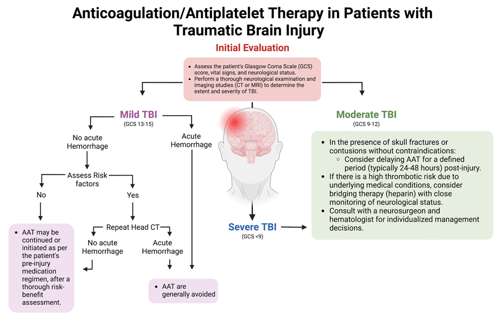 Figure 1: The use of AAT in patients with TBI.
In cases where a patient on AAT sustains a TBI, the risk of bleeding within the brain is heightened due to the ability of these medications to interfere with the blood's ability to clot. Furthermore, the overuse of AAT could also inadvertently result in hypotension, which can reduce cerebral blood flow [12,18]. Consequently, medical professionals must carefully weigh the benefits and risks of continuing or discontinuing these medications during the acute phase of TBI management, and a delicate balance must be struck between preventing further bleeding in the brain and minimizing the risk of clot-related complications [19].
This paper aims to review the literature to better ascertain the appropriate duration of AAT in the context of traumatic brain bleeds. We aim to offer an extensive overview of the current literature pertaining to the ideal duration for administering AAT following traumatic SAH, as well as in cases of acute or chronic SDH. Furthermore, we will explore the optimal duration for antiplatelet therapy and its implications for patient care in skull fractures, cerebral contusions, and DAI. We will also address the impact of AAT on prosthetic valves and stents and its influence on bleeding time and platelet aggregation. Our discussion will extend to the potential future implications for research concerning the utility of AAT in the context of traumatic brain bleeds, emphasizing the existing limitations within the current body of research on this topic.
Researchers for years have strived to elucidate and support the specific decision-making process involved in determining whether a patient should continue AAT following a traumatic SAH [20]. This is a topic of growing prevalence, and a substantial body of research has established connections between the administration of warfarin, an oral anticoagulant, and an increased risk of ICH, inviting further research on the topic [21]. Furthermore, antiplatelet medications, such as aspirin, have been studied for the role they play in preventing inflammation or aneurysm rupture, as depicted in Figure 2.
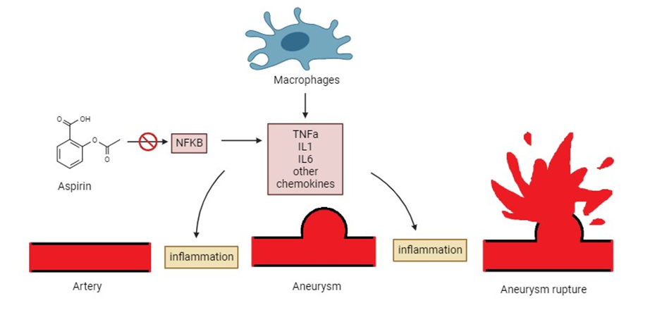 Figure 2: The role of aspirin in patients with SAH.
The optimal timeframe for initiating oral anticoagulation therapy depends on numerous personalized patient factors. Taking this into account, Yan-Huang Li, et al. proposed a straightforward three-step model for determining the ideal timing of AAT administration [20].
The first step of this model focuses on evaluating risk scores for thromboembolism and ICH, which can prove to be a complicated process. For instance, one study by Vivien H Lee, et al. established a simple scoring system known as the HAIR score (Hunt and Hess score, age, intraventricular hemorrhage, re-bleed) [22]. This scoring system, ranging from 0 to 8, offers insight into the various risks associated with SAH and was formulated based on data from 400 patients suffering from traumatic SAH and acute and chronic SDH. The study found that a higher HAIR score was associated with increased hospital mortality among their patients [22]. Another study examined the aneurysmal rupture risk scores in patients with SAH in a large prospective study involving a cohort of 319 patients. Surprisingly, this team of researchers concluded that the aneurysmal risk scores did not correlate with a patient’s increased risk for mortality or intracranial bleeding [23]. This discrepancy between studies calls for more research in the development of models that accurately predict a patient’s risk assessment in order to draw better conclusions regarding the duration of AAT.
The Optimal Timing for AAT in SAH and SDHA retrospective study by Yana Puckett aimed to determine the best timing for resuming AAT in 256 patients with TBI between 2009 and 2012. The study reported that resuming AAT between 7 and 9.5 days after TBI may lead to fewer adverse events when compared to previous recommendations of 3-10 days. The study analyzed patient metrics, such as coagulation studies, type of TBI, treatment, and AAT resumption timing, and it evaluated patient outcomes during a six-month follow-up. Rates of complications were also assessed, including mortality, myocardial infarction, stroke, re-bleeding, venous thromboembolism, and pneumonia. Adverse events were least observed (10%) in the group restarting AAT between 7-14 days, while the group not resuming AAT had the highest rate of adverse events (68.8%). One limitation of this study was low statistical power due to follow-up limitations and a patient cohort with shorter hospital observation periods and more benign brain injuries [17]. Further research should focus on patients with more severe TBIs with longer durations of hospitalization.
Another review article by Jochen A. Sembill concluded that physicians typically initiated AAT as early as one week for patients without significant risk factors like atrial fibrillation or a history of thromboembolism [24]. For patients with other relevant medical histories, the timing of re-initiation was adjusted based on individual risk factors. On the other hand, a survey of multiple neurosurgeons by Yan Xu et al. revealed a lack of consensus regarding the timing of re-initiating therapy, with 40% of physicians preferring to recommence treatment between 14 days and 3 months after the initial injury [25]. In conclusion, the literature offers varying recommendations, with some favoring re-initiation of therapy within one week, others suggesting a 1-2 week delay, and still others advocating for even longer intervals exceeding one month [17,24-29].
Lastly, in terms of how long AAT should be administered, one study by Hormuzdiyar H Dasenbrock, et al. found that long-term AAT was associated with several significant benefits, including shorter hospital stays, reduced risk of venous thrombotic events, and a decreased likelihood of cardiac complications [30]. Similarly, another study concluded that the long-term administration of this therapy (>2 weeks) substantially lowered the overall incidence of delayed cerebral ischemia, particularly when it was concurrently applied with endovascular treatment [31]. Moreover, it was associated with improved patient outcomes and reduced mortality rates. Conversely, a retrospective review study conducted by Ben King, et al. examined the guidelines for resuming anticoagulant therapy and demonstrated the potential risks of hemorrhage and thrombosis [32]. Consequently, these divergent findings suggest that there is an identifiable optimal timing for resuming therapy that falls within the current window for re-initiation.
The administration of AAT following skull fractures, frontal and temporal contusions, and DAI is a topic of ongoing research and clinical debate. However, the absence of high-powered research, such as randomized control trials, limits the availability of evidence-based clinical recommendations in this domain [19]. The decision regarding the management of AAT administration is intricate and multifactorial, encompassing considerations such as the patient's overall health, the severity and location of the injury, and the risk of thromboembolic or hemorrhagic events. Consequently, the approach to managing these patient cohorts relies heavily on a physician's empirical evaluation and clinical experience. This reliance on clinical judgment may contribute to the absence of established protocols for determining the optimal timeframe for resuming AAT in patients following a TBI [14,17,19]. Therefore, there is an imperative need to optimize the timing of AAT administration to enhance patient outcomes.
AAT Indications for Minor TBI and Skull FracturesThe current literature presents a range of recommendations concerning the duration of AAT following TBI. For instance, in cases of minor skull fractures or minor TBI with negative Computerized Tomography (CT) images, there is typically no recommended reversal of AAT. Instead, it is advised to monitor the patient in the intensive care unit and perform repeat scans to rule out any potential bleeding [13,19]. If the subsequent scans do not reveal any bleeding, the resumption of AAT may be considered within a matter of days to weeks, contingent upon other clinical and radiological assessments. However, the optimal duration of AAT for patients with skull fractures remains a topic of ongoing research. This highlights the need for further clinical trials and consensus guidelines to provide clearer recommendations to healthcare practitioners managing these complex cases [16].
AAT Indications for Severe TBI, Cerebral Contusions, and DAIIn cases involving severe TBIs characterized by extensive contusions or DAI, the potential benefits of AAT must be carefully weighed against the associated bleeding risks. This decision often requires multidisciplinary collaboration among neurologists, neurosurgeons, and hematologists to optimize patient outcomes while minimizing complications. However, rapid AAT reversal is recommended in patients with ICH, with platelet transfusion being the suggested approach for reversing aspirin or clopidogrel therapy [13, 19]. In cases of warfarin-induced coagulopathies, effective treatment can typically be achieved with fresh frozen plasma and intravenous vitamin K. However, the use of recombinant activated factor VII in warfarin-associated traumatic ICH remains uncertain, mainly due to limited clinical benefits, cost considerations, and safety concerns in trauma cases [13].
Albrecht, et al. conducted a retrospective investigation into the risk of adverse outcomes in TBI patients hospitalized for one year. They compared those who resumed warfarin therapy to those who did not, revealing a reduced risk of thrombotic events (RR, 0.77; 95% CI, 0.67–0.88) and hemorrhagic or ischemic strokes (RR, 0.83; 95% CI, 0.72–0.96) associated with warfarin resumption. However, warfarin resumption was also linked to an increased risk of hemorrhagic events (RR, 1.51; 95% CI, 1.29–1.78). These findings suggest an overall net benefit in resuming anticoagulation for most patients [33].
Another study, which included 3355 participants, reviewed the outcomes related to neurological deterioration or progression of hemorrhagic TBI on repeat head CT scans in patients who received anticoagulation within 60 days post-injury. The median administration time for anticoagulation after injury was found to be 9 days, and there were no indications of neurological decline attributable to the administration of anticoagulation; however, 6 patients exhibited an advancement of hemorrhagic TBI in follow-up head CT scans. Furthermore, multiple logistic regression analysis revealed that patients aged 65 and older were significantly linked to the advancement of hemorrhagic TBI following therapeutic anticoagulation (odds ratio, 15.2; 95% confidence interval, 1.1-212.7; P=0.04) [34].
Tykocki and Guzek, et al. also reported a relatively low risk of hemorrhagic complications in the early resumption of antithrombotic therapy within 3-17.5 days after TBI. Furthermore, their findings indicate that the risk of such complications may be even lower when non-vitamin K antagonist oral anticoagulants are used, compared to vitamin K antagonists [35].
AAT therapy is also indicated for use in traumatic brain bleeds for patients with prosthetic valves and stents. For instance, one study by Makkar, et al. found that clopidogrel and aspirin therapy significantly reduced thrombus formation in ex vivo porcine models with deployed nitinol stents in external arteriovenous shunts. The study investigated the acute actions of these regimens and found that a combination of clopidogrel and aspirin demonstrated significant effects for reducing acute thrombus formation over the deployed stents, even at low doses. Aspirin showed synergistic effects when combined with clopidogrel, enhancing its anti-thrombotic effects, but models treated with aspirin alone had almost complete obliteration of the stent by the thrombus. Similarly, pigs treated with clopidogrel alone had an insignificant level of diminished thrombosis unless given in relatively high doses when compared to the combined treatment regimen [36].
In another study involving a rabbit model, the combined regimen was assessed in ex vivo heart valves and compared with the acute effects of warfarin. The study found that clopidogrel and aspirin therapy resulted in a greater reduction of thrombus over the heart valves than warfarin or control groups [37].
Similarly, in a different experiment involving baboons, Harker, et al. examined the efficacy of clopidogrel and aspirin in preventing platelet depositions on externally placed arteriovenous shunts with stainless steel stents. The study examined the cumulative effect of clopidogrel in inhibiting platelet depositions over six days on the hypercoagulable stent; it studied low doses of the clopidogrel and examined the additive effects of aspirin when combined with clopidogrel. The study also investigated heparin in an ex vivo placed stent, both alone and in combination with clopidogrel. The study demonstrated that heparin did not significantly reduce platelet depositions over the placed stent unless it was combined with clopidogrel. The study also found that the combination of these regimens did not significantly increase the bleeding time when compared to clopidogrel alone [38].
McKellar, et al. also contrasted the effects of clopidogrel and aspirin with dalteparin in preventing thrombus formation over various periods of time. The study involved a post-mortem analysis of thrombus size over implanted aortic valves. The researchers studied swine for 30 days divided into three treatment groups: 1. combined treatment with clopidogrel and aspirin, 2. single treatment with clopidogrel, aspirin, or dalteparin, or 3. non-treated group. Swine receiving Dual Antiplatelet Therapy (DAPT) had a drastic reduction in thrombus size in comparison to the control group. Additionally, swine treated with single therapies had a larger thrombus size when compared to the DAPTs, but their effects were deemed to be insignificant. The swine receiving dalteparin had the largest thrombus size compared to the other treatment groups, followed by aspirin and clopidogrel. Dalteparin-treated swine had a thrombus size nearly three times larger than DAPTs. DAPTs were also compared to the control group over 150 days. Mean thrombus size in the aspirin and clopidogrel combined group was significantly smaller than the control group, with a four times greater size reduction. Interestingly, there were no reported deaths due to thromboembolic events in the short-term, long-term, or non-treated group [39]. Hence, it can be concluded that a combination of aspirin and clopidogrel is the most effective treatment for thrombi in prosthetic valves and stents.
The Effects of AATs on Intravascular ThrombosisThe debate regarding AAT also extends to the topic of intravascular thrombosis. For instance, one double-blinded randomized control trial studied two groups of cats receiving either aspirin or clopidogrel to investigate the incidence of Secondary Cardiogenic Arterial Thromboembolism (CATE). The researchers found that clopidogrel-treated cats had a significantly decreased likelihood of recurrent CATE and an increased median time for the event to occur. Additionally, the clopidogrel group was less likely to die due to cardiac causes [40].
The aforementioned study by Harker, et al. also found that although heparin and clopidogrel significantly reduced platelet aggregation on the placed stent, they did not decrease the platelet deposition on a vascular graft when compared to clopidogrel alone. Contrastingly, clopidogrel combined with aspirin diminished graft platelet depositions dramatically [38]. Another study measured the acute Cyclic Flow Reduction (CFR) in the coronary arteries of pigs with induced unstable angina and its relation to clopidogrel and aspirin when used alone or combined. These researchers found that clopidogrel and aspirin together, even at low doses, exhibited rapid and effective action in decreasing the CFR whilst inhibition of platelet aggregation was delayed. In fact, aspirin or clopidogrel alone with the same doses did not show any effects at all [41].
The antithrombotic effects of clopidogrel, ticlopidine, and prasugrel were singly compared in rat models after induction of carotid arterial thrombosis in a different study. The study measured vessel occlusion time after induction of thrombosis and treatment in comparison to the non-treated group. Prasugrel displayed more potent and faster actions, compared to clopidogrel and ticlopidine, in keeping patent vessels and reducing thrombus formation [42]. Moreover, these drugs were examined for their ability to prevent arteriovenous shunt thrombosis in rats; their results were strikingly similar to the previously mentioned study [42,43]. In another experiment involving rat models, aspirin, clopidogrel, enoxaparin, and heparin were compared with each other as single therapies or in combination with aspirin for the inhibition of CFR post-induction of carotid artery thrombosis. This study concluded that clopidogrel combined with aspirin was more effective than enoxaparin and aspirin or heparin and aspirin in the reduction of thrombus formation [44]. Combined therapy with aspirin and clopidogrel is more effective in treating intravascular thrombosis than other combination or stand-alone treatment regimens.
The Effects of AATs on Platelet AggregationClopidogrel substantially inhibits ADP-induced platelet aggregation in a dose-dependent manner; however, it has no effect on collagen-induced aggregation. Contrastingly, aspirin prevents collagen-dependent aggregation in a dose-dependent pattern with a minimal effect on ADP-induced aggregation. A combination of both regimens resulted in a delayed effect on ADP-dependent aggregation without a significant effect on collagen-induced aggregation [41]. On the other hand, prasugrel displayed stronger and faster inhibition of platelet aggregation in both mechanisms, and its inhibitory effects increased when combined with aspirin, even at low doses [45,46].
The Effects of AATs on Bleeding TimeThe aforementioned study by H. J. Daykin, et al. compared bleeding time in association with the administration of AAT alone or in combination with aspirin. The tail bleeding times associated with heparin and enoxaparin were significantly increased at higher doses when compared to aspirin or clopidogrel alone. Combining anticoagulants with aspirin further increased bleeding time in contrast to the combination of clopidogrel and aspirin [44]. Similarly, rivaroxaban had a minor increase in bleeding time compared to clopidogrel with aspirin, but no statistically significant differences were found [47]. Clopidogrel and prasugrel also had no significant difference in the bleeding time [46]. Further research should focus on assessing bleeding time with combination therapies with larger sample sizes.
Ticagrelor, Prasugrel, and ClopidogrelAnother experimental study involved ex vivo and in vivo rat and dog models to compare ticagrelor therapy to clopidogrel and compounds 072 and 105, chemical compounds identical to prasugrel and its active metabolite, respectively. J.J.J. van Giezen, et al. found that ticagrelor has distinguishing chemical properties when compared to both prasugrel and clopidogrel. For instance, ticagrelor is an active compound that does not need to be converted by the liver to exert its effects, unlike thienopyridines. Additionally, the high affinity of ticagrelor saturates active sites and leaves it able to more rapidly reach equilibrium when compared to clopidogrel and prasugrel [48]. Ticagrelor is also capable of reversibly binding to the active site, unlike prasugrel and clopidogrel. Additionally, in vivo and ex vivo studies revealed no significant difference in bleeding time and thrombus formation between ticagrelor and prasugrel [49].
The core concern when administering AAT in cases of traumatic brain bleeds revolves around the balance between potential therapeutic benefits and associated risks. These medications are invaluable for mitigating the danger of thrombotic events such as ischemic strokes. However, their mechanism of action also significantly elevates the risk of further hemorrhagic complications such as increased intracranial pressure, brain herniation, neurological deficits, epilepsy, and cerebral edema [50]. While it is widely accepted that platelets regulate primary hemostasis, the impact of antiplatelet medications on subdural hematomas or subarachnoid hemorrhages remains an area of active investigation [51]. However, antiplatelet use has been associated with an increased risk of acute ICH post-head trauma, albeit with a low risk for delayed ICH [52-54]. Thus, immediate head CT imaging is recommended for all such patients [55]. In clinical scenarios where anticoagulation is crucial for the patient, such as in atrial fibrillation or specific cardiac conditions, the decision to recommence treatment should be more meticulously calculated. Overall, the drugs that are currently in use in the context of traumatic brain bleeds are outlined in Table 1.
Table 1: Drugs commonly used to manage coagulopathy in traumatic brain bleeds.|
Drug |
Mechanism |
Side Effects |
Drug interactions |
Reversal agents |
|
Warfarin |
Vitamin K Antagonist |
Bleeding, Bruising |
NSAIDs, Antibiotics |
Vitamin K, FFP |
|
Dabigatran (Pradaxa) |
Direct thrombin inhibitor |
GI Upset, Bleeding |
Antiplatelets |
Idarucizumab |
|
Rivaroxaban (Xarelto) |
Factor Xa inhibitor |
Bleeding, Liver dysfunction |
NSAIDs, Antiplatelets |
Andexanet alfa |
|
Apixaban (Eliquis) |
Factor Xa inhibitor |
Bleeding, Nausea |
NSAIDs, Antiplatelets |
Andexanet alfa |
|
Heparin |
Antithrombin activator |
Bleeding, HIT |
Antiplatelets |
Protamine Sulfate |
|
Enoxaparin (Lovenox) |
Low molecular weight heparin |
Bleeding, Bruising |
NSAIDs, Antiplatelets |
Protamine Sulfate |
|
Clopidogrel (Plavix) |
ADP receptor inhibitor |
Bleeding, GI Upset |
PPIs, Other antiplatelets |
None |
|
Lisinopril |
ACE Inhibitor |
Cough, Elevated blood K+ |
NSAIDs, Diuretics |
None |
|
Amlodipine |
Ca2+ Channel blocker |
Edema, Headache |
Grapefruit juice |
None |
|
Metoprolol |
Beta-Blocker |
Fatigue, Bradycardia |
Antiarrhythmics |
Glucagon |
|
Levetiracetam |
SV2A modulation |
Irritability, Dizziness |
Other CNS depressants |
None |
In patients with multiple comorbidities, polypharmacy is a prevalent issue that further complicates the use of AAT. This polypharmacy environment significantly heightens the potential for drug interactions, making it an area requiring comprehensive study. Certain medications, such as NSAIDs and specific antibiotics, can interact with anticoagulants to exacerbate the risk of bleeding. These drug interactions are not merely additive but can often produce synergistic effects, amplifying the risks associated with each medication [56].
As noted earlier, optimal duration for which AAT should be administered in the context of traumatic brain bleeds remains controversial. Majeed et al. concluded that the optimal timing for restarting anticoagulation should be between 10 and 30 weeks after the intracranial bleeding (55% had ICH), based on the high risk of recurrent hemorrhage in the first 10 weeks that decreased over time, and a rise in ischemic complications beyond 30 weeks [57]. Meanwhile, Park et al. proposed that OAC be reinitiated after 2 weeks to avoid the overriding effect of intracranial hemorrhage recurrence on the prevention of systemic thromboembolism and ischemic stroke [58]. Current practice is largely empirical and there is a need for prospective, randomized trials that compare short-term vs long-term therapy to determine which provides an ideal risk-to-benefit ratio.
While current AAT has proven efficacious in various settings, their limitations, and associated risks in the context of traumatic brain hemorrhage illustrate the need for innovative pharmacological strategies. The development of drugs with targeted mechanisms of action that provide anticoagulant benefits without increasing the risk of hemorrhage is of paramount importance. Promising therapeutics include Novel Oral Anticoagulants (NOACs), next-generation antiplatelet agents, antihypertensive medications, antiepileptics, and statins.
NOACs such as dabigatran etexilate mesylate, rivaroxaban, and apixaban represent a significant leap forward. A 2022 meta-analysis revealed that Direct Oral Anticoagulants (DOACs) cut the risk of ICH by nearly half compared to Vitamin K antagonists, with dabigatran 110 mg identified as the safest DOAC in terms of ICH risk [59-61]. Another study from 2020 found that despite DOACs being reversed at nearly half the rate of warfarin, patients with traumatic ICH on warfarin had higher 6-month mortality rates, suggesting a potential survival advantage for DOACs in this population [62]. As such, NOACs are increasingly becoming first-line options for stroke prevention in atrial fibrillation patients at high risk for ICH [63].
Next-generation antiplatelet agents, such as cangrelor and ticagrelor, hold significant promise in the management of patients who have experienced traumatic brain bleeds [64]. Unlike traditional antiplatelet drugs like clopidogrel, these newer agents offer more targeted and controllable platelet inhibition. For example, cangrelor is an intravenous P2Y12 inhibitor with rapid onset and offset of action, providing clinicians with the ability to quickly titrate its effects [65]. Ticagrelor, on the other hand, is an oral agent that also acts on the P2Y12 receptor but offers the benefit of reversibility [66]. These features balance the need for antithrombotic efficacy with a minimized risk of further hemorrhagic complications. This adaptability offers clinicians greater flexibility in tailoring therapy to individual patient needs, which can be particularly beneficial in scenarios requiring surgical intervention or when there is a high risk of recurrent hemorrhage.
The utilization of antihypertensive agents like ACE inhibitors, calcium channel blockers, and beta-blockers, although somewhat controversial, offers another strategy to mitigate the risk of recurrent bleeds by maintaining optimal blood pressure levels. Antiepileptic medications, such as levetiracetam, may be utilized prophylactically to prevent seizures, which can elevate intracranial pressure and potentially exacerbate bleeding [67]. While not directly aimed at reducing the risk of bleeding, some clinicians consider the use of statins for their neuroprotective effects, although further research is needed to validate their efficacy in this context [68]. Lifestyle modifications also serve as adjunctive strategies and should be monitored rigorously. These include smoking cessation, alcohol limitation, and dietary changes aimed at vascular health.
To refine our understanding of optimal anticoagulation duration in the context of traumatic brain bleed, a comprehensive, multifaceted approach is essential. This would serve to elevate the quality of evidence, allowing clinicians to deliver evidence-based care that maximizes therapeutic benefits while minimizing adverse outcomes. Strategies for achieving this involve the design of rigorous randomized clinical trials, longitudinal cohort studies, meta-analyses, standardization of outcome metrics, and patient risk stratification.
Randomized clinical trials remain the gold standard in clinical research, providing robust evidence on the efficacy and safety of interventions. However, longitudinal cohort studies offer complementary insights into longer-term outcomes and potential late complications associated with varying treatment durations. Such studies could provide critical information on the longitudinal safety and efficacy of anticoagulant regimens in brain hemorrhage patients. Additionally, meta-analyses, which synthesize data from multiple independent studies, offer more reliable estimates of treatment effects which is valuable when individual studies yield small or conflicting results. Outcome standardization is another pivotal aspect. Clear definitions of what constitutes a successful outcome such as clot resolution, absence of rebleeding, or reduced morbidity would facilitate comparability across studies.
A notable deficiency in the current state of research is the lack of patient-stratified studies, which would consider individual risk factors like age, comorbidities, and the specific nature of the brain hemorrhage. For instance, anticoagulant therapy poses a unique risk/benefit profile for elderly, stroke-prone patients compared to younger cohorts [59]. Patient stratification could pave the way for more individualized treatment plans, allowing for the optimized use of anticoagulants and antiplatelets while minimizing adverse outcomes.
As the landscape of traumatic brain bleed treatment continues to evolve, the need for comprehensive, comparative research increases. Advancements in personalized medicine offer new avenues for individualized treatment plans, accounting for variables such as drug interactions, genetic predispositions, and co-morbidities. This multi-faceted approach should involve assessing the optimal duration of AAT, as well as evaluating emerging pharmacological alternatives like NOACs and next-generation antiplatelet agents.
While reinitiating appropriate AAT after an interval of cessation to mitigate the risks of ICH is the standard of care in the context of bleeds in TBI, clinicians differ on the timeline and modality of treatment. Various studies demonstrate that reinitiating AAT decreases long-term risks of thrombotic events and ischemic stroke, but this risk must be balanced with the risks of developing ICH if AAT is reinitiated too soon. The timeline for AAT resumption should be based on interdisciplinary risk stratification that takes into consideration patient risk factors and co-morbidities that may predispose to thromboembolic complications of prolonged AAT cessation.
Traumatic brain injury encompasses a spectrum of injuries that can have significant and far-reaching consequences in the short and long term. Achieving an accurate diagnosis and implementing appropriate management strategies, including decisions related to AAT, should be based on the most up-to-date research and customized to meet the specific needs of each patient whilst taking into consideration co-morbidities and clinically complicating factors. The continually evolving body of literature on this subject highlights the critical importance of ongoing research efforts aimed at generating robust evidence to guide the management and treatment of TBIs, especially in the context of continued AAT.
The authors have no conflicts of interest to report.
No

