 Journal of Medical Research and Surgery
PROVIDES A UNIQUE PLATFORM TO PUBLISH ORIGINAL RESEARCH AND REMODEL THE KNOWLEDGE IN THE AREA OF MEDICAL AND SURGERY
Journal of Medical Research and Surgery
PROVIDES A UNIQUE PLATFORM TO PUBLISH ORIGINAL RESEARCH AND REMODEL THE KNOWLEDGE IN THE AREA OF MEDICAL AND SURGERY



 Indexed Articles
Select your language of interest to view the total content in your interested language
Martínez Herrera Alvaro1,2 , Galván Talamantes Yazmín1*, Vega Rivera Felipe3
1Department of Anesthesiology, Hospital Ángeles Lomas, Jesús del Monte, Mexico.
2Department of Intensive Care, Hospital Ángeles Lomas, Jesús del Monte, Mexico.
3Department of Surgery, Hospital Ángeles Lomas, Jesús del Monte, Mexico.
Correspondence to: Galván Talamantes Yazmín, Department of Anesthesiology, Hospital Ángeles Lomas, Jesús del Monte, Mexico. E-mail: mherreraalvaro@outlook.com
Received date: June 01, 2025; Accepted date: June 10, 2025; Published date: June 17, 2025
Citation: Alvaro MH, Yazmín GT, Felipe VR. Transversus Abdominis Plane (TAP) Block as Anesthetic Technique in a High-Risk Patient: A Case Report. J Med Res Surg. 2025;6(3):88-91. doi: 10.52916/jmrs254178
Copyright: ©2025 Alvaro MH, et al. This is an open-access article distributed under the terms of the Creative Commons Attribution License, which permits unrestricted use, distribution and reproduction in any medium, provided the original author and source are credited.
The TAP is a safe and easy handle technique due to use of ultrasonography. TAP has been described for postoperative analgesia in abdominal surgery, since it produces a reversible block of sensitive peripheral nerves on the lower abdominal wall.
We present a male patient in his 80s with a high cardiovascular and pulmonary risk, anticoagulant therapy who required emergency surgical management. Anesthetic management was performed by a TAP block and sedation, surgery went without complications and a suitable anesthetic status, providing an alternative with multiple benefits related to patient's safety, especially in those with high risk.
Transversus Abdominis Plane (TAP) Block, Incarcerated hernia, Ultrasound, Anesthetic technique.
Transversus Abdominis Plane (TAP) block was first introduced by Rafi in 2001, as a landmark-guided technique via the triangle of Petit [1]. It involves the injection of a local solution between the internal oblique muscle and transversus abdominis muscle, interfascial plane through thoracolumbar nerves from T6 to L1 run, providing sensory innervation to the anterolateral abdominal wall, which, blocked with local anesthetics produce somatic blockade of the abdominis and pelvic wall [2]. Currently, ultrasound usage as a tool in regional anesthesia techniques has allowed to replicate its usage and increase its certainty [3]. These qualities have encouraged the use of TAP block as analgesic adjuvant in multiple abdominal surgeries such as hysterectomy, colectomy, prostatectomy, lipoabdominoplasty, hernia repair, laparoscopic hepatectomy and colo-rectal surgery [4-12]. Nevertheless, there are few reports about the usage of TAP block as the only technique for high risk patients, with acute systemic diseases, elderly adults or even with general or neuraxial anesthesia contraindications [13].
An 84-year-old male patient admitted in the emergency room with acute pain in the umbilical area, together with swelling, reddening and fever. He has a medical history of systemic arterial hypertension, a long lasting evolution of Type 2 diabetes, dilated non-ischemic cardiomyopathy, with Left Ventricular Ejection Fraction (LVEF) of 29%, moderate tricuspid insufficiency and mitral from moderate to severe, permanent auricular fibrillation (CHADS2-VASc 5), with permanent pacemaker, Smoking Index (SI) 13.7, Chronic Obstructive Pulmonary Disease (COPD) and cor-pulmonale with systolic pulmonary artery pressure (sPAP): 50 mmHg. Physical examination in semi-Fowler position revealed blood pressure 110/70 mmHg, heart rate 70 bpm with pacemaker rhythm, precordial auscultation with tricuspid and mitral regurgitant murmur. Respiratory rate 20 bpm, saturation 89% with supplemental oxygen via conventional nasal cannula 2 lt/min, physical examination showed a decrease in general respiratory noises, presence of slight murmurs and infrascapular bilateral crepitant stertors predominantly left-sided. Abdomen with acute pain in the umbilical region, with swelling and colour change. Extremities with significant edema in the lower limbs. Computed tomography of the thorax revealed centriacinar emphysema with bilateral pleura leakage, predominantly left, and passive collapse areas in the lower left lobe with air bronchogram. Coagulation time with PT 15.8s, PTT 72.9s and INR 1.3, due to anticoagulation with dabigatran. He was assessed by the general surgery service and diagnosed with incarcerated umbilical hernia and partial bowel obstruction. An ultrasound of the abdominal wall detected an 8.5 mm hernia defect with a sac measuring 2.5 × 3.3 cm containing lipid content and intestinal loops, without any detectable vascular compromise using Doppler (Figure 1).
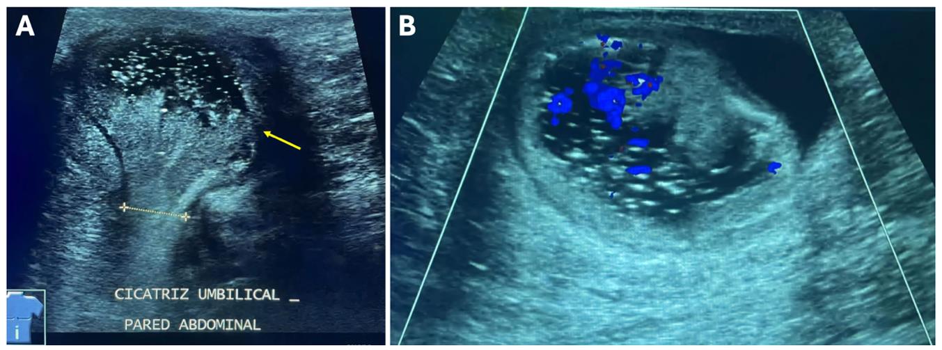 Figure 1: Ultrasound imaging of abdominal wall at umbilical scar. (A): Hernia ring at wall defect (Dotted line) and non-reducible mesenteric and intestinal content (yellow arrow); (B): Vascular compromise of mesenteric and intestinal content of hernia defect with colour doppler.
Immediate surgical resolution is required, with a high anesthetic risk, ASA III, RCRI-Lee of 3 points with 15% risk of high perioperative cardiovascular events and ARISCAT of 80 points with 42% risk of postoperative pulmonary complications, thus, an anesthetic plan with TAP block and intravenous sedation, was decided. The procedure was performed bilaterally with aseptic technique in supine decubitus position and semi-seated at 30 degrees. The site between iliac crest and last costal margin was marked, using the technique described by Hebbard [14] and a posterior approach by means of a lineal catheter of 13-6 MHz was carried out, placing the transducer in transverse plane, above iliac crest at anterior axillary line level, three muscular groups were identified: external and internal oblique muscles and transversus abdominis muscle, as well as intraperitoneal structures. 60 mg of lidocaine at 2% was infiltrated at the site of puncture, using an axon-type needle straight tip 20 gauges with 9 cm length, identifying tip placement between the internal oblique muscle and transversus abdominis muscle, hydro dissection was observed when 3 ml of saline solution 0.9% were injected, locating the target. Afterwards, a total of 15 ml of 0.5% bupivacaine were injected, verifying absence by aspiration of intravascular injection (Figure 2).
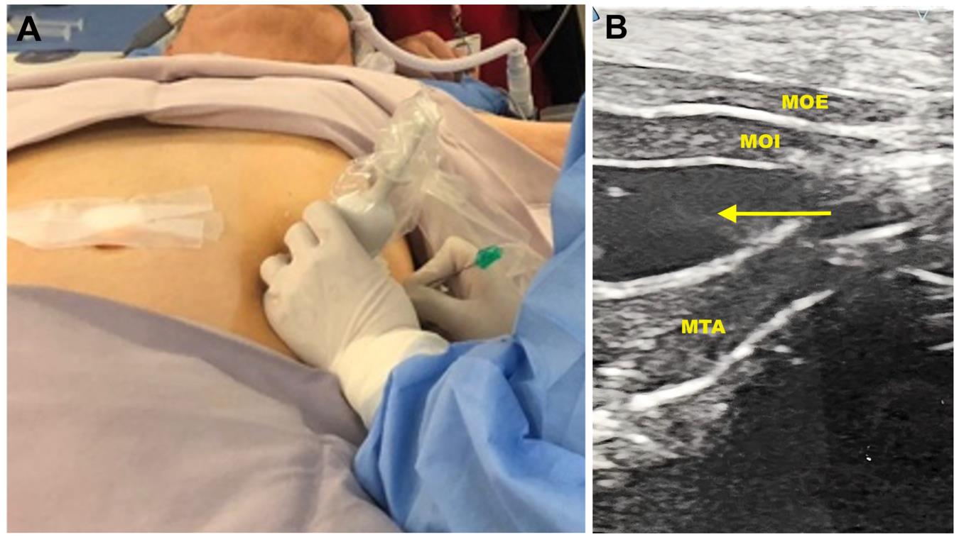 Figure 2: TAP block placement technique. (A): Patient in supine decubitus position, ultrasound transducer perpendicularly to anterior axillary line, between iliac crest and costal margin, and needle insertion in the plane; (B): Identification of MOE, MOI, MTA, and peritoneum via posterior approach; Hydro dissection and local anesthetic infiltration are noted (Yellow arrow).
For anesthetic approach, the patient received supplementary oxygen with nasal cannula 50 lts/min and 50% FiO2, maintaining saturation by pulsi oximetry above 94%. Before surgery, dexmedetomidine in a total dose of 1mcg/Kg was infiltrated intravenously for 20 minutes. Pain was assessed using a verbal rating scale (where 0 meant no pain and 10 severe pain), also, sedation punctuation score (1 fully awake to 3 deep sleep) was recorded. Surgical procedure was carried out according to previous plan, skin dissection, exposure of subcutaneous cellular tissue and hernia sac, then, releasing and shortening of the bowel to the abdominal cavity, in addition to resection of the sac. Primary closure was performed in the peritoneum and aponeurotic defect, by fascia overlap with Mayo´s repair without complications, it lasted 30 minutes with a pain score 0 and sedation score 2. Pain was assessed during post-anesthetic care unit admission at 15, 30 and 45 minutes, maintaining a score of 0.
Evidence reported in literature describes TAP block as a technique that provides postoperative analgesia in abdominal surgery, decreases usage of postoperative opioids in order to improve analgesia combined with other drugs or even, as rescue therapy when other drugs fail [15,16]. Few cases have described TAP block as the sole anesthetic technique for surgeries like inguinal hernia repair, abscess wall drainage, open gastrostomy, colectomy, ileostomy and drainage of hematoma of the rectus abdominis muscle [13,17-20]. TAP blocks provide somatic analgesia of the abdominal wall, including the parietal peritoneum, depending largely on its interfascial extension and, therefore, it does not generate visceral block of the celiac plexus. Several systems classify the TAP blocks into 4, according to the spinal nerves involved.3 In this case, a posterior TAP block was chosen as it blocks anterior and lateral cutaneous branches and is the place of superficial injection for aponeurosis of the lumbar square. Moreover, evidence published by Yoshiyama et al., compared lateral and posterior TAP block in gynecologic laparoscopic surgery and reported a higher analgesic effect in posterior approach [21]. Likewise, Abdallah et al., reviewed results of 12 controlled randomized trials, assessing both approaches (lateral vs posterior), in patients with transversal lower abdominal incisions, showing a decrease in morphine consumption postoperatively with the posterior approach and offering prolonged analgesia [22]. Additionally, data obtained in healthy volunteers studied by magnetic resonance imaging described that posterior approach leads to a greater spread an diffusion even reaching paravertebral spaces form T5 to L [23]. In compromised patients, general and neuraxial anesthesia have also shown several degrees of myocardial depression and vasodilation with adverse effects on circulation.
In this case, anesthetic technique was chosen considering the close relationship between sympathetic and somatic innervation near neuroaxis, as well as its peripheral distance. Besides, few experience is available related with neuraxial anesthesia in patients treated with dabigatran, and any advice about it is based on expert's opinion and its pharmacokinetic knowledge. Dabigatran is a selective thrombin inhibitor, its peak effect is between 30 minutes and 2 hours after ingestion, it is renally excreted in more than 80%. Usage of neuraxial/regional anesthesia in anticoagulated patients treated with dabigatran should be careful and considered individually; a lapse of 34 hours (two half-lives) between the last prophylactic dose and puncture or catheter management, is recommended; this interval could be increased from 72 to 90 hours in patients with creatinine depuration between 30 and 49 ml/min. In case of doubt of any residual effect of dabigatran, it is suggested to use diluted thrombin time (TTd), since prothrombin time (PT) and INR are not correlated with dabigatran plasmatic levels [24]. Activated Partial Thromboplastin Time (aPTT) lengthens non-linearly according to dose, offering qualitative information about its activity in emergencies.
Idarucizumab, a monoclonal antibody that joins dabigatran and reverses its anticoagulant effect has been accepted by the FDA since 2015; although it is not yet available in México [24]. TAP block has been classified among the ones with low risk for bleeding by the European Society of Anesthesiology and Intensive Care as well as the European Society of Regional Anaesthesia [25]. Thus, it provides a safety profile and represents a valuable tool in analgesia and multimodal anesthesia with single-dose techniques and continuous infiltrations [26]. New “deposit” formulations that allow constant release of the encapsulated Local Analgesic (LA) in a biodegradable layer, have been proposed, hence, lengthening its effect for more than 72 hours after the single-dose. This gradual release has demonstrated a safety margin, equal to or higher than conventional LA [27,28]. This formulations could be a valuable tool to control postoperative pain, providing some benefits as higher patient satisfaction and less hospital stay [29]. For example, patients undergoing rotator cuff repair surgery, under brachial plexus block using these formulations, have reported a functional improvement within the first 48 hours, less pain, sleep quality and satisfaction with analgesia [30]. However, these results have not been reproduced in other studies, that do not make any difference related to opioids’ consumption using conventional bupivacaine versus liposomal bupivacaine [31-33]. Even with the introduction of these local anesthetic formulas and with the evidence available, their routine usage is not supported due to the non-meaningful benefit, and the high cost; up to 100 times [28]. So, more high quality and low bias studies are required [27].Interfascial blocks of the abdominis wall represent an excellent option in high-risk patients, however, more studies are required to fully understand their potential as the only anesthetic technique in abdominal wall surgeries. Additionally, excellent communication with the surgical team, knowledge of the technique’s advantages and disadvantages as well as a careful patient’s choice and proper surgical methods are needed, before using a TAP block as anesthetic technique.


