 Journal of Medical Research and Surgery
PROVIDES A UNIQUE PLATFORM TO PUBLISH ORIGINAL RESEARCH AND REMODEL THE KNOWLEDGE IN THE AREA OF MEDICAL AND SURGERY
Journal of Medical Research and Surgery
PROVIDES A UNIQUE PLATFORM TO PUBLISH ORIGINAL RESEARCH AND REMODEL THE KNOWLEDGE IN THE AREA OF MEDICAL AND SURGERY
Journal of Medical Research and Surgery
PROVIDES A UNIQUE PLATFORM TO PUBLISH ORIGINAL RESEARCH AND REMODEL THE KNOWLEDGE IN THE AREA OF MEDICAL AND SURGERY
Journal of Medical Research and Surgery
PROVIDES A UNIQUE PLATFORM TO PUBLISH ORIGINAL RESEARCH AND REMODEL THE KNOWLEDGE IN THE AREA OF MEDICAL AND SURGERY


 Indexed Articles
Select your language of interest to view the total content in your interested language
Yussra Khattri1* , Ashok Kumar1, Danial Khalid Siddiqui1, Bushra Shamim1, Javaria Parwez2
1Department of Radiology, Liaquat National Hospital, Karachi, Pakistan.
2Department of Histopathology, Liaquat National Hospital, Karachi, Pakistan.
Correspondence to: Yussra Khattri, Department of Radiology, Liaquat National Hospital, Karachi, Pakistan.
Received date: January 02, 2025; Accepted date: January 14, 2025; Published date: January 21, 2025
Citation: Khattri Y, Kumar A, Siddiqui DK, et al. Atypical Features of Medulloblastoma in an Adult Patient on MRI: A Rare Case Report. J Med Res Surg. 2025;6(1):1-3. doi: 10.52916/jmrs254156
Copyright: ©2025 Khattri Y, et al. This is an open-access article distributed under the terms of the Creative Commons Attribution License, which permits unrestricted use, distribution and reproduction in any medium, provided the original author and source are credited.
Introduction: Medulloblastoma, a malignant tumor typically located in the posterior cranial fossa, predominantly affects children under the age of 15. It is considered rare in adults. Here, we present an exceptionally uncommon instance of a 35-year-old woman who received a diagnosis of a medulloblastoma located in the cerebellum. Case-Presentation: We report a case involving a 35-year-old female who exhibited symptoms of vomiting and experienced headaches on the right side of her head. The imaging results revealed an unusual mass that was concluded as medulloblastoma on histopathology situated in the right cerebellar hemisphere that extended into the left cerebellar hemisphere. Discussion: The imaging findings indicate the presence of an atypical mass located within the right cerebellar hemisphere, which extends into the left cerebellar hemisphere.This case presents a significant medical concern Further evaluation and management would be necessary. This may include additional imaging studies (such as MRI with contrast), a biopsy to determine the nature of the mass (benign vs. malignant), and consultation with neurosurgeons and oncologists to determine the best course of treatment, which could involve surgery, radiation therapy, chemotherapy, or a combination of these approaches. Conclusion: Medulloblastoma should be considered as a potential diagnosis when evaluating a cerebellar mass. It is important to bear in mind that clinical and radiological characteristics may differ from those typically observed in medulloblastomas found in infants.
Adult medulloblastoma, Atypical features, Cerebellum, MRI, Tumor.
Medulloblastoma is the predominant intracranial tumor in childhood, making up a quarter of all pediatric intracranial tumors and one-third of all neoplasms located in the posterior fossa in children [1,2]. It manifests as a solid, uniformly enhancing mass located in the midline of the posterior fossa. As a Primitive Neuroectodermal Tumor (PNET), it predominantly affects the cerebellum and fourth ventricle [2]. The characteristic clinical and radiological presentation known as "typical" medulloblastoma is not frequently observed in clinical practice. Unusual features may encompass the development of cysts, irregular enhancement patterns, extension through the foramina of the fourth ventricle, manifestation as a tumor in the cerebellopontine angle, and the presence of calcifications. We present a case with an atypical medulloblastoma located in the right cerebellar hemisphere, extending into the left cerebellum [1-3].
We present a case involving a 35-year-old woman who has been experiencing progressively worsening headaches, mainly on the right side, along with bouts of vomiting for the past month. Upon physical examination, the patient was fully alert. Further assessment revealed no signs of cranial nerve dysfunction or any elements suggesting the presence of a tumor outside the skull. The brain MRI displayed a lesion with low signal on t1 and high signal on t2, which exhibited enhancement after contrast (Figure 1). This lesion was associated with surrounding perilesional edema, causing compression on the adjacent parenchyma and a slight elevation of the ipsilateral tentorium cerebelli. There was also herniation of the cerebellar tonsil through the foramen magnum, leading to compression of the medulla. Additionally, compression of the fourth ventricle was observed, resulting in obstructive hydrocephalus affecting the lateral and third ventricles. On the post- contrast study, a branching pattern of enhancement was noted in the right cerebellar hemisphere, extending towards the left cerebellar hemisphere. Spectroscopy indicated the presence of an enhancing mass, characterized by a significantly elevated choline peak, reduced NAA peak, and a choline/creatine ratio ranging from 2.27 to 3.0.
All of these findings are indicative of a high-grade tumor located in the cerebellar hemisphere. The patient underwent surgery, during which histopathology revealed the presence of neoplastic lesion composed of sheets of densely packed cells with hyperchromatic and pleomorphic nuclei with brisk mitotic activity.extensive infiltration int the cerebellar parenchyma and glial tissue is noted representing a high-grade medulloblastoma. she is now under radiotherapy.
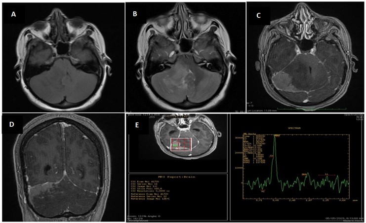 Figure 1: The Atypical features of medulloblastoma on MRI. A): Lesion low signal on t1; B): High signal on t2; C) and D): (axial and coronal): post contrast enhancement. This lesion was associated with surrounding perilesional edema, causing compression on the adjacent parenchyma and a slight elevation of the ipsilateral tentorium cerebelli; E): Spectroscopy indicated the presence of an enhancing mass, characterized by a significantly elevated choline peak, reduced NAA peak, and a choline/creatine ratio ranging from 2.27 to 3.0.
Medulloblastoma is an extremely aggressive tumor that primarily originates from the cerebellar vermis, situated in the roof of the fourth ventricle. The debate over its cellular origin has persisted. Some earlier research posited that medulloblastoma stems from the specific cerebellar granule neuron precursors, which form a germinal center in the rhombic lip at the anterior part of the fourth ventricle [4]. In adults, this tumor more frequently manifests in the cerebellar hemisphere, deviating from the midline vermis involvement more commonly seen in pediatric cases [4,5].
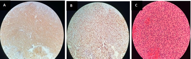 Figure 2: On histopathology. A): The histopathological examination identified a neoplastic lesion characterized by densely clustered cells exhibiting hyperchromatic
and pleomorphic nuclei, accompanied by a high rate of mitotic activity. Additionally, there was substantial infiltration observed within the cerebellar parenchyma;
B): on reaction to synaptophysin; C): Ki 67 proliferative index.
Figure 2: On histopathology. A): The histopathological examination identified a neoplastic lesion characterized by densely clustered cells exhibiting hyperchromatic
and pleomorphic nuclei, accompanied by a high rate of mitotic activity. Additionally, there was substantial infiltration observed within the cerebellar parenchyma;
B): on reaction to synaptophysin; C): Ki 67 proliferative index.The incidence rate in children is nearly 6.0 cases per million population, while in adults, it is less than 0.6 cases per million population. Consequently, the likelihood of this tumor occurring in children is approximately tenfold higher than in adults. Additionally, males have a higher incidence rate of medulloblastoma compared to females [6,7] in our institute we presented with the case of 35 years old adult female.
Patients afflicted with medulloblastoma typically seek medical attention when they exhibit clinical indications of heightened intracranial pressure, often stemming from obstruction of cerebrospinal fluid flow or cerebellar dysfunction. The most prevalent initial symptoms include headaches, nausea, vomiting, and blurred vision. Funduscopic examination may reveal papilledema. As the disease advances and the tumor infiltrates the brainstem, cranial nerves become increasingly affected [1-4] our pt presented with complain of progressively worsening headaches, predominantly on the right side, along with episodes of vomiting for the past month.For children, the presence of a cerebellar mass with low intensity on T1-weighted MRI, alongside hyperdensity on CT scans, strongly indicates medulloblastoma. In adults, MRI findings exhibit a broader range [8]. Tumors appear hypo or isointense relative to the cortex on T1-weighted images, and on T2-weighted images, they can appear hypo, iso, or hyperintense (Figure 2). Contrast-enhanced T1- weighted images in adults reveal varying degrees of enhancement and heterogeneity [3,4]. Typical magnetic resonance images of medulloblastoma display low signal intensity on T1- weighted images and high signal intensity on T2-weighted images. Following the administration of gadolinium, there is pronounced enhancement observed [1]. Atypical imaging characteristics in adult medulloblastoma may be attributed to distinct tumour pathology patterns compared to pediatric cases. Hydrocephalus is also a common feature associated with medulloblastoma [4].
In the presence of calcification, cystic or necrotic areas, poorly defined boundaries, and a lack of enhancement that resembles an extra-axial mass, these characteristics are considered atypical for medulloblastoma in the first decade of life. These features are similar to our case, where the tumor is located in the right cerebellar hemisphere and extends into the left cerebellum [1,2].
The most prevalent tumors found in the posterior fossa during adulthood are metastasis, haemangioblastoma, ependymal tumors, and medulloblastoma [1]. Histological analysis remains the definitive method for diagnosing these tumors. Initial diagnostic insights for posterior fossa tumors can be obtained through MRI findings. However, distinguishing between haemangioblastoma, medulloblastoma, and ependymal tumors based on these imaging features can be a complex task [6]. In literatureview,Spectroscopy revealed a distinct tumor profile characterized by heightened levels of choline and lipids, a decrease in NAA (N-acetylaspartate), and an elevated choline/ creatine (Cr) ratio.(3) In our patient, spectroscopy indicated an enhancing mass, with a significantly elevated choline peak, reduced NAA peak, and a choline/creatine ratio ranging from 2.27 to 3.0.
Regrettably, specific radiological characteristics that definitively differentiate medulloblastomas from other tumors in the posterior fossa have not yet been established. As a result, relying on the pathomorphology of excised tumors remains the most reliable method to confirm or exclude medulloblastomas. The misdiagnosis rate is notably elevated due to the limited understanding of non-cerebellar vermis medulloblastomas [2,9].
Medulloblastoma should be considered as a potential diagnosis for tumors located in the posterior fossa. While adult Medulloblastoma is a rare occurrence, it generally carries a relatively positive prognosis. The presence of Medulloblastoma in adults is exceptionally uncommon, and establishing a diagnosis can be challenging. Relying solely on imaging techniques is insufficient for definitively diagnosing Medulloblastoma in adults, as the tumor exhibits a wide range of characteristics that can be mistaken for other masses in the posterior fossa. In our specific case, confirmation of the Medulloblastoma diagnosis can only be achieved through histopathological examination.
The authors declare no conflict of interest.
The authors have not received any funding for this paper.

