 Journal of Medical Research and Surgery
PROVIDES A UNIQUE PLATFORM TO PUBLISH ORIGINAL RESEARCH AND REMODEL THE KNOWLEDGE IN THE AREA OF MEDICAL AND SURGERY
Journal of Medical Research and Surgery
PROVIDES A UNIQUE PLATFORM TO PUBLISH ORIGINAL RESEARCH AND REMODEL THE KNOWLEDGE IN THE AREA OF MEDICAL AND SURGERY



 Indexed Articles
Select your language of interest to view the total content in your interested language
Bekir Atik*
Department of Plastic and Reconstructive Surgery, Istanbul Medeniyet University, Istanbul, Turkey.
Correspondence to: Bekir Atik, Department of Plastic and Reconstructive Surgery, Istanbul Medeniyet University, Istanbul, Turkey.
Received date: January 28, 2025; Accepted date: February 25, 2025; Published date: March 04, 2025
Citation: Atik B. Assessment and Evaluation of Microsurgery Training J Med Res Surg. 2025;6(2):36-39. doi: 10.52916/jmrs254164
Copyright: ©2025 Atik B. This is an open-access article distributed under the terms of the Creative Commons Attribution License, which permits unrestricted use, distribution and reproduction in any medium, provided the original author and source are credited.
Background: Microsurgery training is very expensive and complex, and achieving a high level of efficiency in a short time is essential. In this study, a scale was developed to make the courses more effective. Material and Methods: A scale with ranges from Level 1 to Level 10 was created, and each level had to be mastered in order to advance to the next level; processes required to be achieved at each level were listed. When trainees achieved these processes, they could then proceed to the next level. Discussion: Those who passed Level 5 were considered to be successful, and in this group of trainees, 92% were successful. Conclusion: There is a program in all courses. Mostly, a program is divided into course days. In this study, it is stated that the programs should be according to the trainee, not the course.
Microsurgery, Education, Assesment, Training, Standardisation.
Repair of tissues and organs with the help of magnification is called microsurgery. The idea that the vessels are untouchable emerged late in the history of medicine. The idea of tying the vessels with a thread to stop bleeding was recorded first in 1564 by Ambroise Paré. Until then, vessels had been considered untouchable [1]. The idea of vascular repair was first introduced by Hallowell who connected a brachial artery by using two pins and then applying a suture in the shape of a figure eight [2]. However, with the introduction of magnifying devices to the surgical field, the utilization of vascular repair surgery gained momentum. In 1902, Alexis Carrel took the first step towards defining microsurgery training in a laboratory as a discipline with his article entitled “Surgical Technique for Vascular Anastomosis and Organ Transplantation,” and he was awarded the Nobel Prize for his work [1,3]. Microsurgery training has progressed slowly, but has gradually gained more importance over recent years. This training requires special laboratory study, and microsurgery training courses are offered throughout the world [4].
The programs designed for many of these courses are similar to each other, and they involve a preliminary study using plastic models, which is followed by vascular anastomosis and neural coaptation studies on animal models. Each trainee follows the same program on the predetermined days; however, this presents a problem, especially in large trainee groups. Some trainees follow the daily program, while others can proceed to the next step before achieving the previous one. This is dependent both on their individual abilities and whether they have previous experience in microsurgery. The Basic Microsurgical Ranking Scale (BMRS) (Table 1) was defined as the basis for assessing the results of the practices of the trainees to ensure better follow-up, as well as to enable the trainees to be more efficient.
From 2013 to 2016, this scale was applied to a total of 82 trainees in groups consisting of four trainees each in the Microsurgery Laboratory of Istanbul Medeniyet University in Istanbul, Turkey. Of the trainees, 36 were plastic surgeons, 15 were orthopedic surgeons, 19 were general surgeons, six were pediatric surgeons, one was an otorhinolaryngology surgeon, four were urology surgeons, and one was a cardiovascular surgeon. The trainees were given information at the beginning of the program, and target goals were identified, but the studies were not scheduled on specific days or at specific hours. After each trainee was interviewed to assess his or her microsurgical knowledge and experience prior to the course, the trainee-specific programs were determined accordingly. The study steps, including hand-eye coordination, plain models, tubular synthetic models, and live animal artery anastomosis, were determined as the steps of the joint program that were required to be achieved [5,6]. (Figure1-6) The more advanced steps involving the use of live animal models were adapted for each individual trainee.
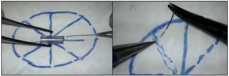 Figure 1: (a): Microscope handling - Level 1; and (b): Plain plastic model study - Level 2.
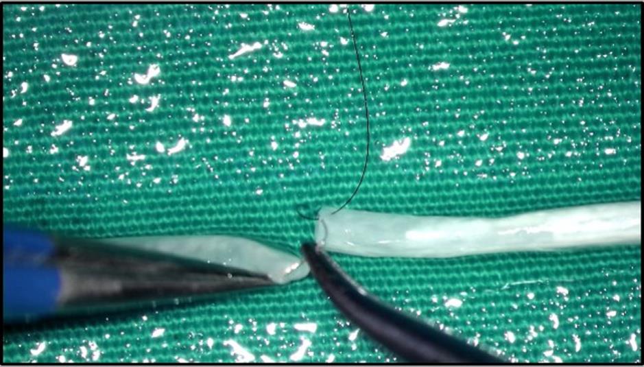 Figure 2: Tubular model study - Level 3
 Figure 3: (a): Rat femoral region dissection - Level 4; and (b): Rat femoral artery anastomosis - Level 5.
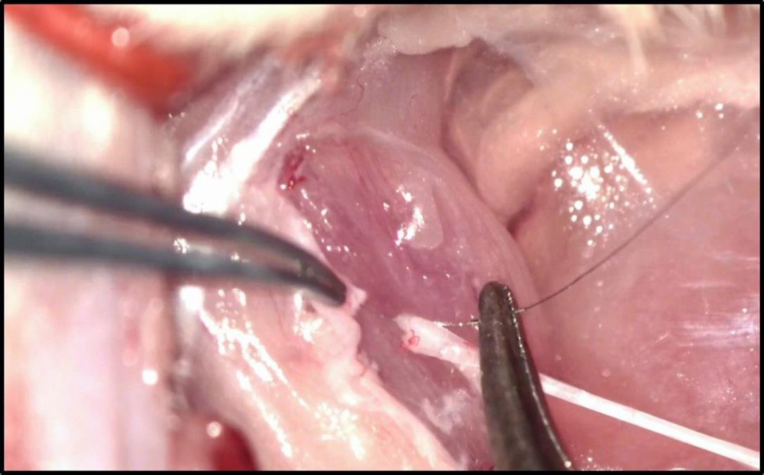 Figure 4: Rat sciatic nerve coaptation (Level 6).
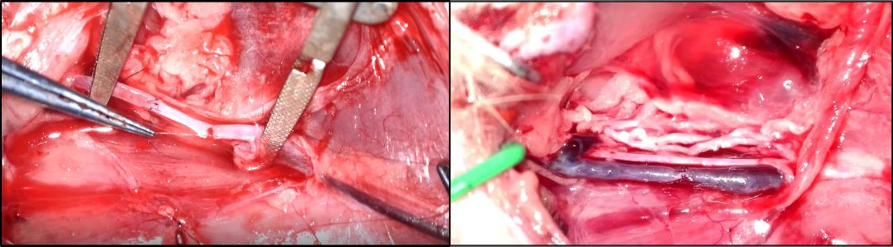 Figure 5: (a): Anastomosis with vein graft, to rat femoral artery - Level 7a; and (b): Rat femoral vein anastomosis - Level 8a.
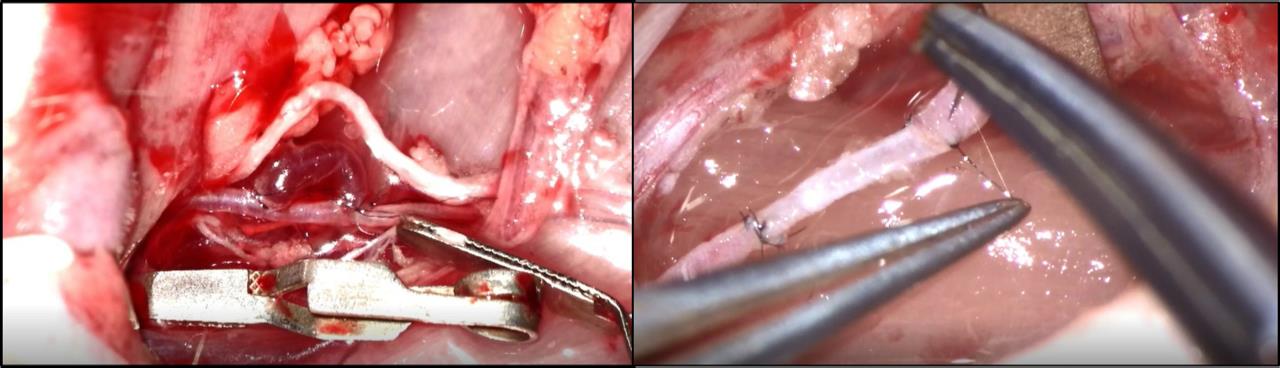 Figure 6: (a): Bypass with a vein graft in femoral artery - Level 9; and (b): Anastomosis with graft vein from the other leg, to rat femoral vein – Level 10.
The study steps were categorized from Level 1 to Level 10; after each application was achieved, the trainee proceeded to the next application (Table1). The trainees who passed Level 5 were considered to be successful. At the end of the course, the levels at which the trainees finished the course were recorded. Also, at the end of the course, the steps which the trainees completed were recorded.
Table 1: Basic Microsurgical Ranging Scale (BMRS).|
Level 1: Microscope handling |
Level 2: Plain plastic model study |
Level 3: Tubular model study |
Level 4: Rat femoral region dissection |
Level 5: Rat femoral artery anastomosis |
Level 6: Rat sciatic nerve coaptation |
|
Familiarization with microscope |
Making incision on the model |
Inserting branula into tubular structures |
Preparing fat flap in the femoral region |
Ability to cut the edge of the femoral artery lumen, and strip its adventitia properly |
Skin incision, and accessing the sciatic nerve |
|
Ability to adjust magnification (zoom) and focus |
Making suture on the model |
Ability to cut the edge of the Lumen vertically and obliquely |
Dissecting the fascia |
Ability to expand the lumen's edge with forceps/needle holders |
Dissection of the nerve from other tissues |
|
Clarifying the object |
Suture knot tying |
Ability to make straight incision (slit) on the body and open a window |
Femoral Nerve dissection |
Placing sutures at equal distance from each other and the lumen's edge. |
Slitting the nerve trunk in a straight line |
|
Binocular vision |
Cutting the suture in desired size |
Placing sutures at equal distance from each other and the lumen's edge |
Femoral Artery -Vein Dissection |
Proper suturing and suture cutting |
Placing epineural sutures at equal distance from the front section. |
|
Coordinated use of both hands with objects (Threading a wire through a bead) |
Placing sutures at equal distance from incision lines |
Ability to control the back wall by using needle holder/forceps |
Dissection of side branches of femoral artery, and their separation and connection. |
Ability to perform "Patency test" |
Turning the nerve and placing epineural sutures on the rear section. |
|
Holding a needle with a needle holder |
Placing sutures at equal distance from each other |
Vessel lumen patency in tubular structures |
Placing Approximator into the femoral artery, and approximation. |
Running anastomosis |
Coaptation without protruding fiber |
If the trainees, one could not complete Level 2, and one could not complete Level 4 and dropped out of the course. Among the remaining trainees, four could not receive a certificate after failing to pass Level 5. Of the remaining 76 trainees, 14 completed the course at Level 10; 10 at Level 9; 30 at Level 8; five at Level 7; 2 at Level 6; and 15 at Level 5. Fifty-eight percent of the trainees planned to perform the vascular anastomosis process in the clinical trials. Eighteen of the trainees attended the course to conduct the dissection process, using the microscope, in order to gain the ability to intervene in possible complications (such as vessels or nerve incisions) as their main objective, in addition to performing the vascular anastomosis process. When those trainees passed Level 5, their studies focused on partial vascular dissection and partial and full neural repair.
The International Coach Federation (ICF) has defined coaching as the process of collaboration between coach and trainee [7]. In microsurgery coaching, the work that needs to be done on each course or training day should be well-organized and optimized. In addition, the planned works should be controllable and measurable. A separate program needs to be determined for each course to be taught and each study to be carried out within the scope of microsurgery, and the subunits in such a scheduled program must be distinctive. In many training courses, other than microsurgical ones, the required processes to be carried out are standardized [1,4,8]. They are known and do not vary depending on the trainee or the course. However, in microsurgery, the processes should vary depending on both the course and the trainee; in some cases, it should be possible to apply several separate programs in a single course. The coach should personalize the program by considering the educational background of the trainee. By using this scale, we determined the steps to be accomplished after Level 5 in accordance with the needs of the trainees.
Microsurgery training is a very difficult training that requires intense concentration. It is important to apply a different program to each trainee and follow up with him/her individually. This is difficult to do in an overcrowded course. Therefore, many centers limit the number of trainees they accept. Microsurgical training courses are held in various countries where successful microsurgeons are trained. However, similar programs are carried out, program steps are grouped into certain days in many of the courses, and the trainees are asked to strictly adhere to these schedules [8].
Most trainees attend the course with some apprehension [9]. The trainee is able to overcome this apprehension by discussion and by learning the relevant techniques, from basic to advanced. After passing the stages, the trainee begins to gain self-confidence. Preparing a tailor-made program for each trainee is important in this regard. If all the trainees do the same thing, this results in the disappointment, and possibly the failure, of some trainees. A trainee who is mentally ready, skillful, or has already been engaged in microsurgery previously will have different needs than the other trainees. At each stage of the course, trainees should be allowed to learn at different steps. This rating scale enables trainees to follow their own agendas and study independently of the other trainees’ programs.
In fact, the courses are conducted in many organizations that provide training for microsurgery, and these often take into consideration the aforementioned issues. However, assessment, evaluation, and standardization are not currently available in the administration of these courses. Standardization of the courses will be made easier by the implementation of this scoring system. Surveys conducted in some courses shed light on how that course will be organized in the future [10].
The scale that we have prepared will facilitate the storage and standardization of data and will also contribute to the design of the courses that will be offered in the future. The administrators of each course can prepare a scale similar to this. This scale has been prepared in accordance with basic microsurgery training requirements. This rating scale can be modified depending on the nature of the course offered. For example, the program can be initiated from Level 5 or above; it can also be designed in such a way as to include a free flap scale, replantation scale, or transplantation scale.
Today, courses taking fewer than five days are conducted globally [11]. In such courses, it is possible to achieve greater efficiency in a shorter time by using this scale before initiating the program to determine the level at which each trainee must begin. The only disadvantage of this scale is that it requires a close follow-up of each trainee. However, receiving the best training from microsurgery courses is only possible when they are conducted with a small number of trainees.
In this course, unlike in other courses, the procedures to be conducted by the trainees were not organized into days. The trainees were prompted to accomplish the steps, and their success was determined based on the steps that they reached by the end of the course. The applied scale was revised and adjusted according to the needs of the individual trainee. We recommend that training center authorities whose centers offer such courses create their own scales.
No.


