 Journal of Medical Research and Surgery
PROVIDES A UNIQUE PLATFORM TO PUBLISH ORIGINAL RESEARCH AND REMODEL THE KNOWLEDGE IN THE AREA OF MEDICAL AND SURGERY
Journal of Medical Research and Surgery
PROVIDES A UNIQUE PLATFORM TO PUBLISH ORIGINAL RESEARCH AND REMODEL THE KNOWLEDGE IN THE AREA OF MEDICAL AND SURGERY
Journal of Medical Research and Surgery
PROVIDES A UNIQUE PLATFORM TO PUBLISH ORIGINAL RESEARCH AND REMODEL THE KNOWLEDGE IN THE AREA OF MEDICAL AND SURGERY
Journal of Medical Research and Surgery
PROVIDES A UNIQUE PLATFORM TO PUBLISH ORIGINAL RESEARCH AND REMODEL THE KNOWLEDGE IN THE AREA OF MEDICAL AND SURGERY


 Indexed Articles
Select your language of interest to view the total content in your interested language
Tatjana Barišić1* , Anđela Azinović2
1Department of Obstetrics and Gynecology, University Clinical Hospital Mostar, Bosnia and Herzegovina.
2Department of Surgery, University Clinical Hospital Mostar, Bosnia and Herzegovina.
Correspondence to: Tatjana Barišić, Department of Obstetrics and Gynecology, University Clinical Hospital Mostar, Bosnia and Herzegovina.
Received date: November 30, 2023; Accepted date: December 07, 2023; Published date: December 14, 2023
Citation: Barišić T, Azinović A. Adnexal Torsion in the Second Trimester of Pregnancy: A Case Report and Review of the Literature. J Med Res Surg. 2023;4(6):118-120. doi: 10.52916/jmrs234123
Copyright: ©2023 Barišić T, et al. This is an open-access article distributed under the terms of the Creative Commons Attribution License, which permits unrestricted
use, distribution and reproduction in any medium, provided the original author and source are credited.
Acute abdomen in pregnancy is one of the biggest diagnostic and therapeutic challenges today. Despite advances in medical technology, the preoperative diagnosis of acute abdominal conditions is still inaccurate. Torsion of the adnexa, which refers to complete or partial rotation of the adnexa, resulting in obstruction of venous and lymphatic reflux in the ovary, is a common gynecological emergency. Torn adnexa affect both the ovary and the fallopian tube, and rarely affect only one of them. Adnexal torsion occurs more frequently in the first and early second trimester than in the third trimester. It is difficult to diagnose in advanced pregnancy because of nonspecific symptoms and signs of acute abdomen as well as ultrasound limitations due to uterine enlargement complicate diagnosis. Late diagnosis and delayed surgical treatment result in ovarian loss and fetal endangerment. It is important to thoroughly evaluate the adnexa at the 1st ultrasound examination in pregnancy and to monitor the observed formations.
Acute abdomen, Adnexal torsion, Pregnancy.
Acute abdomen in pregnancy is one of the biggest diagnostic and therapeutic challenges today. The incidence of acute abdomen during pregnancy is 1 in 500 pregnancies. Despite advances in medical technology, the preoperative diagnosis of acute abdominal conditions is still inaccurate [1,2]. The development of ultrasound diagnostics and antenatal protection has resulted in an increase in the incidence of adnexal tumors in pregnancy, which reaches up to 1% to 2%. These are most often benign formations, of which 80% withdraws spontaneously as the pregnancy progresses. Torsion of the adnexa, which refers to complete or partial rotation of the adnexa, resulting in obstruction of venous and lymphatic reflux in the ovary, is a common gynecological emergency. Torn adnexa affect both the ovary and the fallopian tube, and rarely affect only one of them [1,2]. The risk of adnexal torsion is affected by the size of the adnexal mass. Adnexal torsion occurs more frequently in the first and early second trimester than in the third trimester. It is difficult to diagnose in advanced pregnancy because of nonspecific symptoms and signs of acute abdomen as well as ultrasound limitations due to uterine enlargement complicate diagnosis. Late diagnosis and delayed surgical treatment result in ovarian loss and fetal endangerment [2-4].
We present a case of 21-year-old primipara (G1, P-0, AB-0) who reported to the emergency medicine center in the 19th week of pregnancy due to severe pain in the right lower abdomen, which started 10 h ago, but became unbearable 3 h ago. During the examination, the patient complains of pain in the lower right abdomen, has extremely poor clinical picture, feels exhausted, looks pale, and feels general weakness and nausea, vomited 3 times, with a tendency to worsen. Clinical history revealed that the patient was healthy before the pregnancy, and the regularly controlled pregnancy has been in order so far. Clinical examination verifies markedly painful right abdomen on palpation, temperature of 37.3 Celsius, pulse of 92 Beats per Minute (BPM), respiratory rate of 20 breaths per minute and blood pressure of 120/70 mmHg. Laboratory results on admission: L 9.3, E 4.2, 123 HB, HCT 0346, TRC 140; GUK 6.5, urea 2.1, creatinine 47, CRP 1.8. From urine sediment: L 10-14, abundant bacteria (++), leukocyte esterase (+), proteins (+).
During the gynecological examination, the doctor found neither vaginal discharge, nor bleeding, nor pelvic mass in the pelvis. However, soreness and bodily habitus due to soreness limit the examination. Obstetric ultrasound showed that it was a normal "single pregnancy" with a fetal heart rate of 160/ min, biometrics corresponds to the duration of amenorrhea. TV cervicometry: CL=43 mm, "T" shape. Ultrasound of the abdomen showed approximately 3 cm × 2.3 cm of free fluid along the intestine, i.e. the bottom of the cecum, with echogenic surrounding adipose tissue. Based on all of that, situation was discussed with the surgeon on duty who estimated that it was most likely the case of acute appendicitis and the method of an emergency laparotomy according to Sprengel's was chosen. The operation found an appendix without inflammatory changes and an appendectomy was performed in the usual way. Then a twisted gangrenous tumor of the right ovary with a diameter of approximately 15 cm × 10 cm, right Infundibulopelvic (IP) ligament twisted three times, was found, and a gynecologist who was on duty performed a detorsion and right adnexectomy with a slightly enlarged incision, leaving the cystic structure intact along the abdominal cavity. The procedure of lavage was performed and a drain was set up.
The preparation is sent for PH analysis. The procedure went smoothly. Pathohistological findings showed:
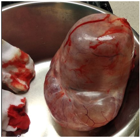 Figure 1: Ovarian tumor-teratoma.
Microscopically, the fallopian tube is free of pathological changes, and inside the ovary, cystic formations lined with cubic to cylindrical epithelium can be seen, while some cystic formations are lined with multilayered squamous epithelium under which numerous skin adnexa can be seen. Among the described cysts, a loose stroma with blood vessels, clusters of siderophages can be seen, and few giant multinuclear cells of the foreign body type in some places. One cut contains the remains of a yellow body. It's a mature teratoma. The postoperative course went smoothly, infusion solutions, analgesic and anticoagulant therapy, dual antibiotic therapy with Clindamycin 3 × 900 mg/5 days and Ceftriaxone 1 g/5 days, Immunoglobulin anti D, since the pregnant woman had a negative blood type, supported by the intravaginal progesterone of 200 mg for the next four weeks. The later course of the pregnancy was orderly and in the 40th week, a healthy female newborn, 3850/56, the optimal Apgar score, was born.
Adnexal torsion refers to the total or partial twisting of the ovary around its vascular axis and can result in vascular compression and necrosis. A total of 13.7% of all episodes of adnexal torsion were found in pregnant women. In a ten-year review of the study, ovarian torsion accounted for 2.7 percent of emergency surgeries and was the fifth most common surgical emergency [5]. Ovarian cysts during pregnancy can be conservatively monitored by observation if diagnosed in the first trimester. The optimal time for surgical intervention is between 16 and 28 weeks of gestation. Urgent surgical intervention may be justified in case of ovarian torsion, rupture of the ovarian cyst or if malignancy is suspected [6]. The diagnosis of ovarian torsion in pregnancy can be made by clinical imaging in combination with color Doppler ultrasound. Treatment options are limited to surgery, laparoscopic approach, or laparotomy. Pregnancy loss is rare and postoperative progesterone supplementation is recommended when the corpus luteum is removed, before 7th to 9th week of gestation [7-9]. The incidence of adnexal masses decreases with advancing gestational age due to spontaneous disappearance of most benign and physiological adnexal masses, but also due to reduced possibility of palpation and ultrasound display of adnexa with advancing pregnancy. Those with a simple cystic appearance can be conservatively monitored by serial ultrasound monitoring. However, they may need an emergency exploratory laparotomy due to rupture, torsion or infarction in as many as 50% of the cases [6]. Ovarian torsion during pregnancy is a rather uncommon complication with high patient morbidity and fetal mortality if not treated immediately. Torsion is more common on the right rather than the left side with an incidence of 3:2.1. The reason is probably that the sigmoid colon restricts the mobility of the left ovary. Ultrasound is a safe, accurate, fast, non-invasive and inexpensive primary method for detecting adnexal torsion. Sonographic findings of adnexal torsion include unilateral ovarian enlargement (>4 cm), uniform peripheral cystic structures, coexistent mass within the affected ovary, free pelvic fluid, lack of arterial or venous flow, and twisted vascular pedicle. The presence of flow at a color Doppler dose does not preclude a torsional diagnosis. Pena et al. [10] reported that 60% of 21 surgically confirmed cases of ovarian torsion were normal on Doppler ultrasound, leading to a delayed diagnosis of up to 2 days. Increased gas in the intestines, small field of vision, obesity, and anatomical changes in late pregnancy can interfere with proper visualization of the adnexa, making it difficult to assess adnexal torsion. With the increase in the number of ultrasound examinations in the first trimester of pregnancy due to screening for aneuploidies, the number of accidentally detected adnexal masses increased. The total estimated incidence of adnexal masses in pregnancy is 2% to 10%, or 1 in 25 to 1 in 8000 pregnancies. Certainly, the induction of ovulation in the treatment of infertility also contributed to an increase in the incidence of adnexal masses in pregnancy. Magnetic Resonance Imaging (MRI) is a safe and ultrasound-complementary method for assessing adnexal masses in pregnancy, especially in the case of unclear ultrasound findings of solid adnexal tumors and fibroids. Color Doppler can only be a complementary method to the morphological ultrasound assessment of adnexal masses in the differentiation of benign from malignant formations [11-13]. If the infundibulopelvic ligament is successfully detorsed, it is common practice to drain the cyst which caused the torsion to prevent recurrence of the torsion. Because a typical first-trimester cyst is a corpus luteum cyst, which supports pregnancy until the placenta is adequately developed at the end of the first trimester, drainage or excision of the cyst can result in pregnancy loss. Therefore, obstetricians choose to treat with additional progesterone to support pregnancy until the placenta develops, usually in the second trimester. Diagnosing ovarian torsion is not easy and is often missed. The signs and symptoms often mimic other neo-obstetric disorders such as appendicitis, pyelonephritis and nephrolithiasis, uterine torsion caused by axial rotation due to the presence of uterine fibroids. We look critically at this case, and we want to emphasize that it is very important to evaluate the adnexa at the 1st ultrasound examination in pregnancy, which in this case was the failure of the gynecologist in primary care, who controlled the pregnancy. We also warn that in the case of an acute abdomen in pregnancy, it is important to think differentially about the torsion of the adnexa in pregnancy, which was missed during the gynecological examination of the abdomen. In our case, the enlarged uterus moved the adnexa and no clear boundaries were found between the right edge of the uterus and the right ovary, so surgeons, after appendix removal, failed to determine whether the mass originated from the right ovary or uterus and only after widening the laparotomy incision, could make a definitive diagnosis of ovarian torsion. Surgery remains the standard method of final diagnosis and treatment.
Teratomas are tumors of germ cell origin that most commonly develop in the gonads. Benign Cystic Teratoma (BCT) is the most common benign ovarian neoplasm, comprising 10% to 15% of all ovarian tumors. It occurs at all stages of life, and most cases are diagnosed between the ages of 20 and 30 [11]. This fact makes it the most common tumor during pregnancy (22% to 40% of all ovarian tumors) [11]. During pregnancy, the risk of complications increases significantly, including rupture, torsion, infection and malignant degeneration. Because BCT tends to remain within the true pelvis, this could lead to dystocia and difficult delivery [11].
It is important to thoroughly evaluate the adnexa at the 1st ultrasound examination in pregnancy and to monitor the observed formations. Any unclear condition of the acute abdomen in any woman with sudden severe pain in the lower abdomen must include differential diagnosis and the suspicion of adnexal torsion. It is always necessary to consult a gynecologist, because it is a question of future fertility, and a successful and timely detortion saves the adnexa and protects fertility.
The authors have no conflicts of interest to report.
No.

