Nicole Dimitra Sarantis1, Evgenia Kalogridaki2, Chrisostomos Sofoudis1*
11st Departement of Obstetrics and Gynecology, General and Maternity Hospital, Elena Venizelou, Greece.
21st Department of Surgery, Breast Unit, General and Maternity Hospital, Elena Venizelou, Greece.
Correspondence to: Chrisostomos Sofoudis, 1st Departement of Obstetrics and Gynecology, General and Maternity Hospital, Elena Venizelou, Greece.
Received date: August 12, 2024; Accepted date: August 22, 2024; Published date: August 30, 2024
Citation: Sarantis ND, Kalogridaki E, Sofoudis C. A Low-Grade Appendiceal Mucinous Neoplasm Mimicking a Hydrosalpinx: A Case Report and Review of the Literature. J Obst Gynecol Surg. 2024;5(1):1-5. doi:10.52916/jogs244038
Copyright: ©2024 Sarantis ND, et al. This is an open-access article distributed under the terms of the Creative Commons Attribution License, which permits unrestricted use,
distribution and reproduction in any medium, provided the original author and source are credited.
Introduction: A Low-Grade Mucinous Neoplasm (LAMN), formerly known as “mucocele” or “cystadenoma,” is a non-invasive and rare appendiceal tumor, representing 0.2-0.7% of all appendix specimens. Case Report: A 31-year-old female patient who was suffering from chronic right lower quadrant abdominal pain was submitted to an exploratory laparotomy for a presumed right hydrosalpinx. A low-grade mucinous neoplasm of the appendix was discovered and the patient underwent an appendectomy. Discussion: A review of the current literature was performed regarding LAMNs. Conclusion: Further evidence is required in order to address the controversial issues of the pre-operative diagnosis of a LAMN, the extent of surgery performed and the questions regarding fertility preservation in young women.
LAMN, Mycocele, Appaendiceal tumors, Hydrosalpinx, Pregnancy, Magnetic Resonance Imaging (MRI), Computed Tomography (CT)
Appendiceal tumors are extremely rare as they account for less than 1% of all cancers and are found at a rate of 0.2-0.3% [1-3]. A Low-Grade Mucinous Neoplasm (LAMN), formerly known as a “mucocele” or a “cystadenoma,” is a non-invasive and rare appendiceal tumor, representing 0.2-0.7% of all appendix specimens [4]. It usually presents as a cystic dilation of the appendix, which is caused by the accumulation of mucin produced by neoplastic cells [5, 6]. In this article, we present a rare case of a young patient with LAMN which was misdiagnosed for a hydrosalpinx and we review the relevant literature. To our knowledge, very few such cases have been described and, therefore, we aim to raise awareness regarding this entity that should be included in the differential diagnosis by gynecologists, general surgeons and radiologists.
In 2024, a 31-year-old female (gravida 0 para 0) was referred to the outpatient gynecologic clinic of our tertiary hospital for evaluation of a presumed right hydrosalpinx, which is a dilated fluid-filled fallopian tube. The patient had been suffering from chronic right lower quadrant abdominal pain in the previous four years. Her medical history was unremarkable other than the fact that she had been submitted to an exploratory laparoscopy for the same complaint in 2020; no hydrosalpinges and no other abdominal pathology had been revealed. In 2021, she was submitted to a Magnetic Resonance Imaging (MRI) scan of the abdomen, which demonstrated a 9.8 × 6.2 × 4.2cm cystic formation, located behind the uterus and in contact with the right ovary and had been presumed to be a hydrosalpinx. For the following three years, she had been monitoring this formation with annual sonograms and MRI scans; the formation had remained unchanged in size and characteristics and no abnormal abdominal lymph nodes had been found. She wished a future pregnancy but would not attempt to get pregnant while the issue of the hydrosalpinx remained unresolved.
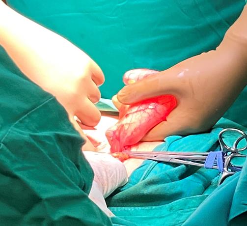 Figure 1: Macroscopic identification of Mucocele.
It was decided that after three years of monitoring she be submitted to an exploratory laparotomy. Intraoperatively, the ovaries and fallopian tubes appeared free of malignancy. There was some free abdominal fluid which was sampled for a cytological examination and was found to be free of abnormal cells. Her appendix, however, was dilated to an approximate size of 10 cm and was obviously the formation that in the sonograms and MRI scans had been mistaken for a hydrosalpinx (Figure 1). An appendectomy was performed by the on call general surgeon, with care not to rupture the appendix (Figures 2, 3).
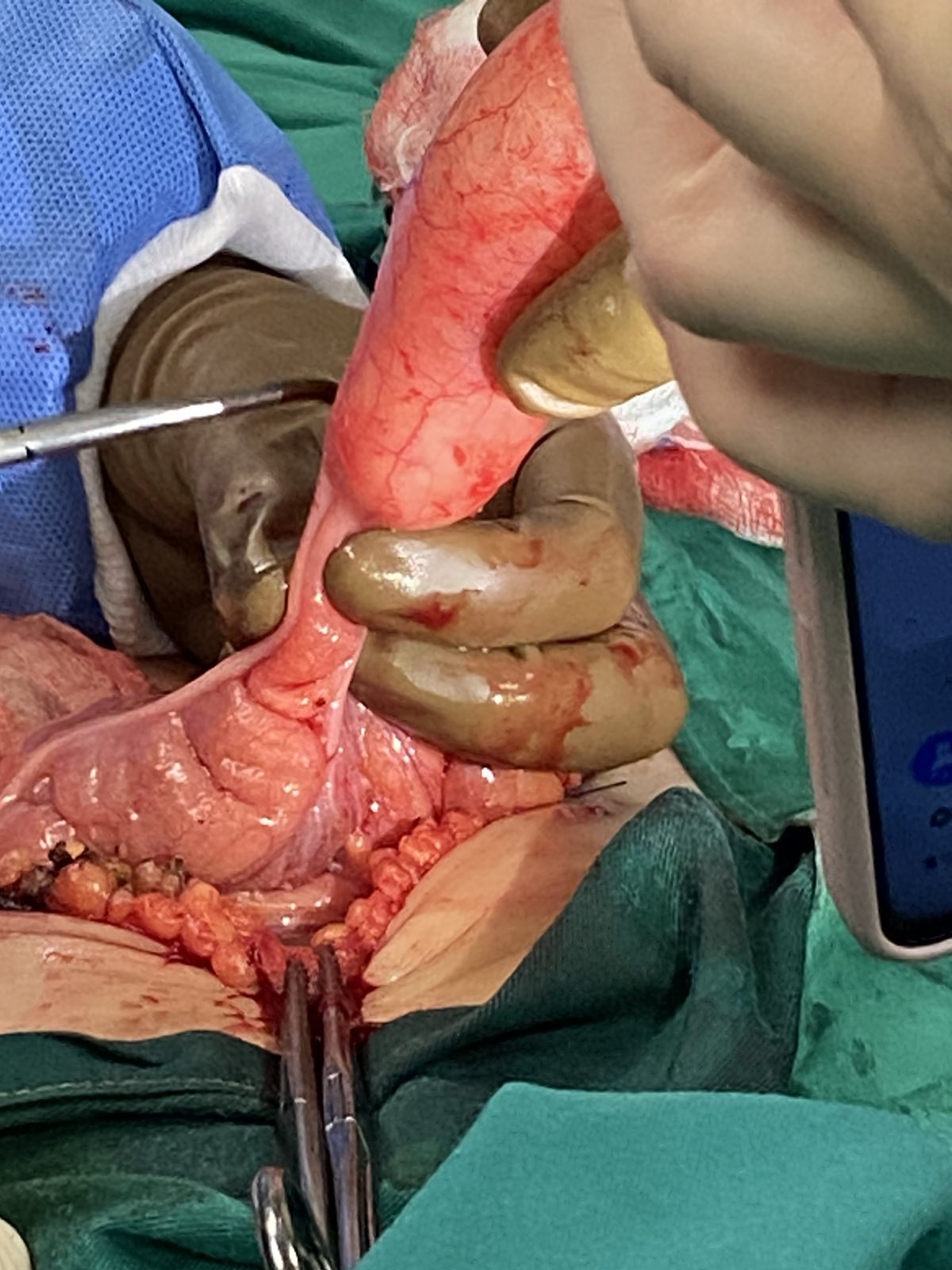 Figure 2: Surgical dissection-Appendectomy.
 Figure 3: Pathological-Specimen.
The peritoneal cavity was carefully examined for abnormal lymph nodes and for mucinous lesions that might have been implanted in it. The surgical specimen was submitted for histological evaluation which revealed a Low-Grade Appendiceal Mucinous Neoplasm (LAMN), stage 3 (according to the 8th edition of ASCC: Cancer staging manual) with clear resection margins. Immunohistologically, it stained positive for CK20 and Desmin. The patient’s postoperative course was uneventful and she was discharged to the care of an oncologist for further follow-up. She is to be monitored every six months and it has not been deemed necessary that she undergo further excisional surgery or chemotherapy.
Appendiceal tumors are extremely rare as they account for less than 1% of all cancers and are found at a rate of 0.2-0.3%, with an annual incidence of six cases per 1000000 people [1-3]. They constitute a heterogeneous group of epithelial and nonepithelial tumors; the group is composed of five different histological subtypes: a) neuroendocrine neoplasms, which are nonepithelial; b) mucinous neoplasms; c) goblet cell adenocarcinomas; d) colonic-type adenocarcinomas; and e) signet ring cell adenocarcinomas; which are all epithelial tumors [2]. Mucinous neoplasms, our patient’s type, constitute one third of all epithelial tumors of the appendix. Unlike our case, where our patient was in her thirties, these tumors are more common in the fifth and sixth decade of life and in women [1,7]. In particular, our patient had a Low Grade Mucinous Neoplasm (LAMN), which is non-invasive and rare, representing 0.2-0.7% of all appendix specimens [4]. According to the 2016 definition established by the Peritoneal Surface Oncology Group (PSOGI), LAMNs are defined as mucinous neoplasms with low-grade cytology and any of the following properties: fibrosis of the submucosa, loss of the lamina propria and muscularis mucosae, a pushing pattern of growth into the wall imparting an expansile or diverticulum of acellular mucin into the wall, or mucin and/or neoplastic mucinous epithelium outside the wall of the appendix [1]. The term “mucocele” has also been used to describe mucinous neoplasms as the appendix becomes dilated due to the accumulation of mucin produced by the neoplastic cells [5,6]. LAMNs were also formerly known as mucinous cystadenomas [8,9].
An appendiceal mucocele can be asymptomatic or it may present with lower abdominal or pelvic pain, a palpable mass, distention, intestinal obstruction, fever, nausea, vomiting, and weight loss. Most commonly it may mimic appendicitis (in 50% of cases) or an ovarian cyst or a hydrosalpinx, which was the case with our patient [1, 4, 6, 8, 10-17]. Rare presentations have also been described; a mucocele mimicking a perforated diverticulitis [18] or presenting with urological symptoms such as hematuria, ureteral obstruction, hydronephrosis [6] or mimicking a subserous uterine fibroid [10].
Diagnosis can be aided by imaging techniques such as an ultrasound, Magnetic Resonance Imaging (MRI), Computed Tomography (CT) and colonoscopy. For instance, an appendiceal mucocele can be distinguished from an uncomplicated appendicitis sonographically by the lack of appendiceal wall thickening of >6 mm [7]. Moreover, a mucocele may appear sonographically as a bottle-like shaped solid appendicular mass sliding over the uterus and ovaries. It may also present with the “onion skin” sonographic sign, which is concentric echogenic layers with septa and fine echoes and which distinguishes a mucocele from an ovarian cyst. In our case, the tubular mass that was apparent in the sonograms was initially misdiagnosed for a hydrosalpinx due to its proximity to the right ovary. Applying pressure with the ultrasound probe (the so-called “split” sign) separates a lesion from the ovary and, thus, may be indicative when a cystic lesion of non-ovarian origin lies in proximity to the ovary [10]. Colonoscopic features that may be helpful in reaching a diagnosis are the presence of a bulbous submucosal lesion of the cecum and the “volcano” sign, which is an appendiceal orifice seen in the center of a firm mound covered by normal mucosa [1,7,15]. On MRI, a mucocele is seen as a well encapsulated cystic mass that appears hyperintense on T2-weighted sequences, as was the case with our patient. On a CT scan, apart from the typical appearance of a cystic mass, calcifications of the cyst wall are considered characteristic of a mucocele (Aleter). Even so, a CT scan is diagnostic only in 50% of cases [10].
It is worth noting that patients with LAMN may have elevated tumor markers including Carcinoembryonic Antigen (CEA), Cancer Antigen-125 (Ca-125) and Carbohydrate Antigen 19-9 (Ca 19-9), which is the case with 56-67% of patients, and, therefore, can be used in monitoring prognosis post-diagnosis [4]. In our case, however, the patient was negative for all tumor markers.
According to the American Joint Committee on Cancer (AJCC) Staging Manual, a 3-tiered approach is used in assigning mucinous neoplasms a grade as follows: G1 means low-grade well-differentiated tumors, G2 means high-grade moderately-differentiated tumors and G3 means high-grade poorly differentiated tumors with signet ring cells present [1]. It is highly important that when examining the specimen, LAMNs, which are by definition low grade, be distinguished from HAMNs (High Grade Mucinous Neoplasms) and mucinous adenocarcinomas, as the latter have worse outcomes, prognosis, recurrence rates and, of course, management; details regarding HAMNs and adenocarcinomas are outside the scope of this article and will only be mentioned briefly in relation to LAMNs. Immunohistology and molecular profiling may facilitate the diagnosis of a LAMN on pathological examination and may aid in distinguishing it from a HAMN. A HAMN is a neoplasm that shows architectural features of a LAMN, has no infiltrative growth or destructive invasion, but shows areas of high-grade cytologic atypia or complexity; these features include enlarged hyperchromatic and pleomorphic nuclei or architectural features such as cibriform architecture, micropapillary features or piling up of epithelial cells. Appendiceal mucinous neoplasms are profusely stained positive for CK20, MUC5AC and DPC4 and negative for CK7 [3, 4]. Both LAMNs and HAMNs frequently show mutations in KRA, GNAS and RNF43. However, in contrast to LAMNs, HAMNs also have mutations in TP53, ATM and APC [19]. Mucinous adenocarcinomas, on the other hand, exhibit features indicative of invasion such as: stromal desmoplasia, frankly infiltrative glands, destructive invasion and the appearance of tumor cells floating in small pools of mucin. Mucinous adenocarcinomas may present along with LAMN or HAMN and thus it is very important for pathologists to examine the appendix on its entirety.
Regarding the staging of LAMNs, tumors confined to the muscularis propria are assigned the T category Tis; pT1 and pT2 are not applied in LAMNs. If acellular mucin or mucinous epithelium is identified beyond the muscularis propria in the subserosa or mesoappendix but does not involve serosal (visceral peritoneal) surface, the tumor is designated as pT3. If the mucinous epithelium or acellular mucin present on serosal surface, then the tumor is classified as pT4a. M1a is applied when there is intraperitoneal acellular mucin but no epithelium is identified. M1b means that there are intraperitoneal mucinous deposits containing epithelium [19]. The concept of N designation is not applicable in appendiceal mucinous tumors. Peritoneal spread of appendiceal mucinous neoplasm typically leads to extensive mucinous deposits, a condition called mucinous carcinoma peritonei or, most commonly known as Pseudomyxoma Peritonei (PMP). The afore-mentioned grading system of appendiceal mucinous neoplasms (G1 through G3) is also applied in PMP cases [19].
If left untreated, the complications of LAMN include intussusception, volvulus, small bowel or ureteral obstruction, rupture and mucinous ascites [10]. Moreover, an untreated localized LAMN can progress to an advanced, metastatic or inoperable disease. An important sequela of LAMN is the afore-mentioned pseudomyxoma peritonei, which is the peritoneal dissemination of mucus-producing neoplastic cells that may cause the development of diffuse intra-abdominal gelatinous ascites. This condition is a rare entity with an incidence of 1 in a million annually and it is usually the result of a ruptured mucinous neoplasm of the appendix that may happen naturally or iatrogenically. It is worth noting, though, that mucinous neoplasms of other structures, such as the ovary, can also lead to a PMP [20].
The management of mucinous neoplasms of the appendix is rather controversial and is often complicated by the fact that these neoplasms may be diagnosed incidentally either during an operation or post-operatively after the specimen has been evaluated histopathologically. The extent of surgical treatment is at the center of the debate. Treatment of a mucocele requires the surgical removal of the appendix with care not to cause rupture and endoperitoneal spillage of neoplasmatic mucinous cells. A simple appendectomy with post-operative surveillance is recommended for localized LAMNs as they very rarely lead to lymph node metastasis [2,9,10]. It is deemed efficient even in cases where the appendectomy margins involve acellular mucin or neoplastic epithelium and in cases where there is acellular mucin on the appendiceal serosal surface without peritoneal dissemination. In these cases, in fact, it has been noted that supplemental right hemicolectomy after the initial surgery does not provide advantages over appendectomy and it may cause the disease to reach the retroperitoneum and may increase the risk of recurrence in the anastomosis line [6, 21]. It is worth mentioning that certain authors have also described a technique called radical appendectomy which involves removing a cuff of the cecum without involving the ileocecal valve [22]. High-grade mucinous neoplasms, on the other hand, may exhibit node involvement; grade 2 in 17% and grade 3 in 72% of cases and, therefore, require that a right hemicolectomy be performed even in localized cases [2].
There is no consensus as to whether a laparoscopic or open approach is preferable for performing an appendectomy so both approaches are acceptable [1, 2]. It has been suggested, however, that a laparoscopic approach may confer a higher risk of iatrogenic rupture of the appendix [8,11,15].
In cases of LAMNs where peritoneal dissemination is discovered upon exploring the abdominal cavity, management requires appendectomy, irrigation, biopsy of peritoneal nodules, peritonectomy and chemotherapy [6]. LAMNs with localized acellular mucinous deposits carry a 4% risk for developing PMP, while those with cellular mucin are at a higher risk of 40% [2]. In cases where there is metastatic disease in the peritoneum and PMP, the current gold standard is aggressive Cytoreductive Surgery (CRS) and application of Hyperthermic Intraperitoneal Chemotherapy (HIPEC). The most commonly used chemotherapeutic agents in HIPEC for mucinous neoplasms are mitomycin C, cisplatin, oxaliplastin and 5-fluorouracil [2,3,19,20]. Complete Resection (CCR) after CRS is defined as no evidence of visible disease; CCR1 means that less than 2.5mm of disease could not be resected, CCR2 means that that there is 2.5-5 mm disease left and CCR3 means there is more than 5mm of visible disease left after surgery [2,3].
As far as systemic chemotherapy is concerned, it is not recommended that it be administered in cases of LAMN and low-grade PMP. On the contrary, systemic chemotherapy is recommended for grade 2 and grade 3 mucinous neoplasms and PMP, either pre or post-operatively. Fluorouracil-based regimens are mostly used. Even in cases of unresectable disease, chemotherapy may provide disease control in more than half of them [2,3].
Surveillance for five to ten years after appendectomy is necessary to monitor for recurrent disease and PMP. It should include annual CT scans and monitoring levels of tumor markers CEA, CA 19-9 and CA-125 [1,3,6]. Localized LAMN has a five-year survival rate of 95%. If, however, it involves extra-appendiceal spread then it drops to 86% and if PMP is developed then the five-year survival rate further drops to 25%. Applying CRS and HIPEC for aggressive disseminated peritoneal disease results in a five-year survival rate of approximately 50% and a ten-year survival rate of approximately 30%. In cases of incomplete cytoreduction, the five-year survival rate drops at 20% [1,3,6].
An issue of importance that should be addressed is the need to preserve fertility in the small subset of young patients that will need to undergo either CRS and HIPEC or systematic chemotherapy as part of their treatment plan. First of all, during CRS ovaries are most often resected as they constitute a common site for neoplastic implants [23]; the bilateral adnexectomy performed during CRS inevitably leads to infertility. Moreover, even if the adnexa are preserved, adhesions formed due to CRS can lead to decreased tubal motility. In addition, the disease itself (PMP) is considered a factor causing infertility [24-26]. Secondly, HIPEC is a rather controversial issue with regards to ovarian function. Older studies describe how HIPEC leads to impaired gonadal and tubal function: a) it may cause direct damage to germ cells and to maturing follicles that are at a chemotherapy-sensitive stage and b) it results in sclerosis throughout the peritoneal cavity; this fibrotic process causes extensive adhesions and, therefore, infertility [25,27]. On the other hand, recent studies prove that achieving a pregnancy through natural conception is feasible after HIPEC [28-31]. In fact, a meta-analysis conducted by Papageorgiou et al. described the achieving of a spontaneous pregnancy after HIPEC in 16 cases [30]. The rate, however, of spontaneous pregnancies is very hard to determine, due to the rarity of these cases and the lack of data, and is postulated to be low [27, 30, 31]. Thirdly, systematic chemotherapy, as is well known, causes premature ovarian failure, especially in women of older age and women treated with alkylating agents [25,30,31].
It has become, therefore, imperative that fertility-preserving strategies be developed for this rare sub-group of women with mucinous appendiceal neoplasms in need of CRS and HIPEC/systematic chemotherapy. As far as the operation is concerned, it has been proposed by certain authors that the ovaries, tubes and uterus be spared if the ovaries appear macroscopically normal during surgery, using strict criteria [28]. Also, it has been suggested that GnRH agonists be administered prior and during chemotherapy in order to protect maturing follicles from reaching a chemo-sensitive stage. Furthermore, some authors have suggested performing an experimental “therapeutic” laparoscopy with the goal to postpone CRS and HIPEC until after child-bearing in cases of low-grade PMP. This technique entails performing a laparoscopy, appendectomy, copious irrigation and washout of the pelvis with stripping of mucinous disease off the ovarian surfaces [26]. Despite the above-mentioned alternative treatment plans, the standard of care with regard to preserving fertility is to counsel patients to undergo ovarian cryopreservation (or embryo cryopreservation) in order to achieve a future pregnancy through reproductive-assisted techniques. Cryopreservation of ovarian tissue is not usually recommended as it has the potential of re-introducing disease into the abdomen with the ovarian transplant [27,30,31].
In conclusion, our case and review of the literature serve to highlight three main points. Firstly, a LAMN presenting in a young woman is very rare and, therefore, it is imperative that gynecologists, general surgeons, as well as radiologists, be aware of it; it should be included in the differential diagnosis pre-operatively so that, if confirmed intra-operatively, the optimal steps be performed in order to safely remove it. Secondly, there is a debate regarding the extent of resection and surgical treatment; it is of utmost importance that patients avoid a potentially unnecessary second operation that might not confer any benefit at all. Finally, the issue of fertility preservation in younger women with mucinous tumors needs to be addressed; a question arises with regards as to how aggressively we should manage a tumor that might not be aggressive.
All authors declare no conflicts of interest with respect to this manuscript.
No

